My Patient Tools
Save what matters. Prepare your questions. Return to the evidence.
Send my saved information
Prepare an email containing your saved Anchors or Consultation notes. Choose a recipient and press Send in your own email application.
Highlight a passage and select Save to My Anchors. Use saved passages to prepare your consultation.
My Anchors
My Consultation
Select the evidence you want to discuss with your doctor.
Beta: Your information is saved in this browser. Export important notes. Avoid storing private information on shared devices.
Initialising Patient Tools...
01 | PD-L1 Negative. Still Responds: What Are We Missing?
A patient-led investigation into the clinical evidence, the biology behind an unanswered question, and what it means for patients with cholangiocarcinoma.
In CheckMate 067, a clinical trial involving patients with advanced melanoma, 41.3% of patients classified as PD-L1 negative responded to nivolumab alone.
The trial used a 5% tumour-cell staining threshold to define PD-L1 positivity.
That was the observation.
What the result did not explain was why those patients responded.
I came to this question as a patient, not a scientist. Having experienced Stage IV cholangiocarcinoma and a complete response to immunotherapy, I wanted to understand what had made my response possible. More importantly, I wanted to understand what the science could reveal for patients who were still searching for a treatment opportunity.
The number that stopped me was 41.3%.
These were patients whose tumours had been classified as PD-L1 negative under the trial's testing threshold. Yet approximately four in ten experienced an objective response to a medicine that blocks the PD1 immune checkpoint.
How could that happen?
The result contained two important findings. The first was that nivolumab produced measurable responses in these patients. The second was that patients classified as PD-L1 negative were among those who responded.
The trial had demonstrated both findings.
What it had not explained was why these patients responded despite their negative classification.
That was the question I wanted to investigate.
To understand why this matters, we first need to understand something about the immune system and how cancer can interfere with it.
Inside the tumour
Visualise a tumour as a growing collection of cells. Each cell is its own tiny Cell City, carrying instructions and operating systems that determine how it functions.
Around those cells are blood vessels, supporting tissue and immune cells, all interacting within the same environment.
Among the immune cells are T cells. Some are capable of recognising and attacking cancer cells.
But the immune system must also protect healthy tissue from unnecessary attack. It needs controls that restrain its own activity.
One of those controls is a receptor called PD1, found on activated T cells and certain other immune cells.
Think of PD1 as a brake receiver.
Another protein, PD-L1, can connect to that receiver and deliver a signal telling the T cell to apply the brake.
PD-L1 can be expressed by healthy cells, cancer cells and other cells within the tumour environment.
In our Cell City picture, imagine a cell displaying a message that says, "Stand down, maintenance underway."
That protective system serves an important purpose in normal biology. But cancer cells can exploit the same mechanism to weaken an immune attack.
Medicines such as nivolumab, known as Nivo, and pembrolizumab, known as Keytruda, bind to PD1 and prevent inhibitory signals from PD-L1 and another protein, PD-L2, from engaging that receptor.
They do not directly attack cancer cells in the way chemotherapy does.
Instead, they remove one restraint on immune cells that may already be capable of attacking.
Preventing those proteins from applying the PD1 brake does not guarantee a response. The immune system must still recognise the cancer and have the capacity to act.
But it creates an opportunity.
Now return to CheckMate 067.
The trial classified tumours as PD-L1 positive when at least 5% of the assessed tumour cells met the staining threshold. Below that threshold, they were classified as negative.
That classification did not necessarily mean PD-L1 was entirely absent. It did not measure PD-L2. And it did not tell us everything happening between the immune system and the tumour.
Yet patients classified as negative responded.
These were melanoma patients, not cholangiocarcinoma patients. Their response rate cannot be transferred to bile duct cancer.
But the biological question was not confined to melanoma.
What had the test measured, what had it left unseen, and what might explain the responses?
That became my investigation.
The question a study sets out to answer may not be the most important question its results uncover.
02 | How Allison and Honjo Changed Cancer Treatment Forever
James Allison and Tasuku Honjo helped establish a fundamentally different approach to cancer treatment: instead of attacking cancer cells directly, treatment could release restraints on the patient's own immune system.
The CheckMate 067 result could not have occurred without an earlier change in how scientists understood the immune system.
For decades, cancer treatment had largely been organised around attacking cancer directly.
Surgery removed cancerous tissue. Radiation damaged cancer cells. Chemotherapy interfered with their ability to survive and divide.
But researchers were exploring another possibility.
What if the immune system already possessed the ability to attack cancer, but something was preventing it from doing so effectively?
James Allison investigated CTLA-4, a protein involved in regulating T-cell activation.
Tasuku Honjo's laboratory identified PD1, another receptor that helps control immune-cell activity.
These were not two names for the same mechanism.
They were two different controls operating within the immune response.
How does a T cell know what to attack?
Think of a T cell as a member of our body's security team.
Before it can launch an effective attack, it needs to recognise information about a possible threat and receive the appropriate activation signals.
Other immune cells, particularly dendritic cells, can collect pieces of abnormal proteins and display them to T cells.
Think of this as the security team receiving information about a possible threat.
But recognising that information is not always enough to activate the T cell fully.
Additional signals help determine whether it should respond.
One of its important activation receptors is called CD28.
Think of CD28 as a receiver for the additional message telling the T cell it has permission to activate.
CTLA-4 competes for the same signalling partners that activate CD28.
When CTLA-4 takes up those connections, less of the activating signal reaches CD28. The T cell may therefore activate less strongly or fail to become fully activated.
Think of it as the security team recognising a threat but having its authorisation to respond restricted.
Blocking CTLA-4 can help restore that activation signal.
PD1 acts differently.
It can apply another brake to T cells that have already been activated, including when they encounter cancer cells within a tumour.
In our Cell City picture, CTLA-4 helps regulate whether the security team becomes fully activated.
PD1 can slow an activated team while it is attempting to carry out its work.
Both controls serve legitimate purposes.
Without sufficient restraints, the immune system can damage healthy tissue.
But cancer can exploit those restraints.
Two scientists, two treatment opportunities
Allison's work helped establish that blocking CTLA-4 could strengthen anti-tumour immunity.
This contributed to the development of ipilimumab, often called Ipi.
Research following Honjo's discovery established the therapeutic potential of blocking PD1.
This contributed to medicines including nivolumab and pembrolizumab.
In 2018, Allison and Honjo shared the Nobel Prize in Physiology or Medicine for discoveries that transformed cancer immunotherapy.
Their work changed the question being asked about cancer treatment.
Instead of asking only how to damage or destroy cancer cells, researchers could investigate whether the patient's own immune system was being prevented from doing a job it might otherwise perform.
But releasing a brake does not guarantee that the immune system will recognise and eliminate the cancer.
Think of a vehicle.
Releasing the brakes permits movement, but it does not supply the engine, fuel or direction.
Similarly, blocking CTLA-4 may help strengthen T-cell activation, while blocking PD1 may release restraints on T cells already participating in an immune response.
Neither guarantees success.
That distinction becomes important later in this investigation, particularly when considering why a combination of CTLA-4 and PD1 blockade is not biologically identical to PD1 treatment alone.
For now, it brought me back to the original observation.
If patients classified as PD-L1 negative were responding to nivolumab, what restraint was the medicine releasing?
Was the test failing to describe something important about the biology?
Or were other mechanisms involved?
The responses had occurred.
Understanding why required looking beyond the treatment itself.
Releasing the brake creates an opportunity. Understanding what enables the immune system to use that opportunity is the next question.
02 (II) | How Unanswered Cancer Questions Can Become Half-Truths
A scientific study can answer the question it was designed to investigate while revealing another question that remains unresolved.
When that unanswered question could affect a patient's survival opportunity, it must not disappear as the evidence moves into clinical practice.
This is where the problem becomes larger than PD-L1.
Scientific findings travel through research papers, conferences, clinical guidelines, hospitals and eventually into conversations between doctors and patients.
During that journey, complicated findings must be summarised.
That is necessary. Nobody can carry every detail of every study into every consultation.
But something important can happen during that process.
The answered part of the research becomes easier to communicate. The unanswered part may gradually receive less attention.
A finding that was originally limited to a particular experiment, patient population or measurement can begin to sound like a complete biological conclusion.
What began as "We don't yet know" can eventually be heard as "It cannot happen."
Those statements are not equivalent.
I have attended a cancer conference where experienced oncologists, surgeons and radiologists were confronted with the question of why patients classified as PD-L1 negative could respond to PD1 blockade.
The answer was essentially that we did not know.
I do not regard that answer as a failure of the clinicians.
Acknowledging uncertainty is essential to good science and medicine.
What concerns me is what happens next.
Does the unanswered question remain visible? Is it investigated further? Does it continue to influence how we interpret the evidence?
Or does the clinical result travel forward while the biological question is left behind?
A scientist may reasonably ask whether a study answered the question it was designed to answer.
A clinician must ask what the available evidence supports for the patient sitting in front of them.
But the patient has another question, because the patient carries the consequence.
What did the evidence reveal that remains unexplained, and could understanding it change my opportunity for survival?
These questions do not compete with one another. They examine different responsibilities arising from the same evidence.
I am not asking scientists to claim what they have not established, or clinicians to ignore the limitations of research.
I am asking that uncertainty be described accurately.
When something has not been established, we should say so.
When something has been demonstrated, we should not allow a different unanswered question to erase it.
And when an observation could change a patient's opportunity to cure or extend survival, it deserves to remain part of the investigation until it is resolved, disproven or shown not to matter.
The unanswered question must travel with the answer when the patient's opportunity may depend on it.
03 | Was CheckMate 067 an Isolated Result?
CheckMate 067 was not the only melanoma trial documenting responses among patients classified as PD-L1 negative.
Other studies found responses with nivolumab, pembrolizumab and checkpoint combinations.
The finding was repeated, although the studies did not establish why those responses occurred.
One unexpected result can be an unusual observation that does not appear again under further examination.
So I went looking for the same finding in other trials.
CheckMate 066 provided another piece of evidence.
Among patients receiving nivolumab whose PD-L1 status was negative or indeterminate, the reported objective response rate was 33.1%.
The classification was not identical to CheckMate 067, but the finding was relevant.
Patients without a positive baseline PD-L1 classification were responding.
Then came KEYNOTE-006, which investigated pembrolizumab against ipilimumab.
In the reported PD-L1-negative subgroup, approximately 25% responded to pembrolizumab, compared with 13% receiving ipilimumab.
Now the observation extended beyond nivolumab.
Two different medicines acting on the PD1 receptor had produced responses among patients classified as PD-L1 negative.
What happened when two checkpoints were blocked?
In CheckMate 067, nivolumab combined with ipilimumab produced an objective response in 54.8% of the PD-L1-negative subgroup.
CheckMate 069 also reported substantial responses among patients receiving the combination.
These results must be understood carefully.
Combining PD1 and CTLA-4 blockade changes more than one immune control, so the responses cannot be attributed to PD1 blockade alone.
KEYNOTE-029 added another observation.
In its reported PD-L1-negative subgroup, 50.0% achieved an objective response and 33.3% achieved a complete response.
These percentages came from particular patient groups, treatments and assessment methods.
They are not general predictions for everyone whose tumour tests PD-L1 negative.
But they introduce another question that will become important when we reach cholangiocarcinoma.
If blocking PD1 alone can produce responses, and blocking PD1 together with CTLA-4 can also produce responses, why should the results of a chemotherapy and PD1 combination trial be treated as the final answer about either of those other treatment approaches?
They are different biological interventions.
We should not assume that their effects are interchangeable.
Response is not the same as cure
The language of clinical trials matters.
An objective response includes both partial and complete responses.
A partial response generally requires at least a 30% reduction in the measured target lesions under RECIST 1.1.
A complete response means the disease meets the trial's criteria for disappearance of detectable cancer on assessment.
It does not prove that every malignant cell has been eliminated.
How long the response lasts is another question.
These distinctions matter because I do not want patients to mistake a response rate for a cure rate.
But neither should we overlook what was actually demonstrated.
Across different studies, patients classified as PD-L1 negative experienced measurable responses to PD1 inhibitors.
Some patients receiving checkpoint combinations achieved complete responses.
The studies involved different populations, treatment regimens and testing methods.
Their percentages cannot be compared as though they came from a single experiment.
Nevertheless, the central observation survived.
A negative baseline PD-L1 classification did not establish that checkpoint treatment could not produce a response.
The investigation could now move forward.
Instead of repeatedly asking whether these patients responded, we could ask what their original test results had failed to reveal.
The trials confirmed the responses. They did not establish which biological mechanisms made those responses possible.
04 | What Does a PD-L1-Negative Biopsy Actually Tell Us?
A PD-L1 test measures expression in the tissue examined, using a particular method and threshold.
It does not provide a complete account of immune activity throughout the tumour or across time.
The trial evidence had established that patients classified as PD-L1 negative could respond to PD1 treatment.
I now wanted to understand the test itself.
When a patient is told that their cancer is PD-L1 negative, what has actually been measured?
A pathologist examines a sample of tumour tissue and uses a laboratory staining technique called immunohistochemistry, or IHC, to detect PD-L1 protein.
Depending on the cancer type and test, the result may be expressed using different scoring methods.
One method is the Tumour Proportion Score, or TPS, which considers the proportion of viable tumour cells showing the required PD-L1 staining.
Another is the Combined Positive Score, or CPS, which incorporates staining in certain immune cells as well as tumour cells.
These are not interchangeable measurements.
The test also needs a threshold that determines how the result is classified.
In CheckMate 067, the threshold was 5% tumour-cell PD-L1 expression.
A result below that threshold was classified as negative.
But below 5% does not necessarily mean zero.
And even if no PD-L1 staining is detected in the particular sample examined, that does not prove that PD-L1 is absent everywhere in the tumour.
The limits of the sample
Imagine standing in a city and taking a photograph of one street.
The photograph may accurately show what was happening on that street at that moment.
But it cannot tell you everything happening in the surrounding neighbourhoods, and it cannot show what happened yesterday.
A tumour biopsy has a similar limitation.
Different regions of a tumour may contain different populations of cancer cells, immune cells and supporting tissue.
PD-L1 expression may vary between those regions.
A biopsy taken from one part of a tumour may therefore provide a different picture from tissue taken elsewhere.
There is another important distinction.
PD-L1 can be expressed by cancer cells and by other cells in the tumour environment.
A scoring method that focuses on tumour cells may not describe the full pattern of PD-L1 expression in surrounding immune cells.
And PD-L1 is not the only protein capable of engaging the PD1 receptor.
PD-L2 can also deliver an inhibitory signal through PD1.
A PD-L1 test does not measure PD-L2.
This does not establish that PD-L2 caused the responses in CheckMate 067.
But it means the negative PD-L1 classification cannot exclude PD-L2 as a possible contributor.
The test may be accurate while still being incomplete as a description of the entire checkpoint environment.
What should the patient know?
This discovery changes how I would want a patient to understand their pathology report.
A label of positive or negative is not always enough.
I would want to know which assay was used, which scoring method was applied, the numerical result where available, the threshold used, and where and when the tissue was collected.
I would also want to distinguish between a result of zero, a low result, an inconclusive result and a test that was never performed.
These are different findings.
Not reported does not mean negative.
And negative does not necessarily mean that the relevant protein was entirely absent.
This is not an argument that every patient with every cancer must undergo PD-L1 testing.
It is an argument for preserving and accurately interpreting the information whenever the test is performed.
But the biopsy has one further limitation that deserves investigation.
It measures tissue collected at a particular moment.
What if the tumour's immune environment changes?
The biopsy is a photograph. The immune response is a movie.
05 | What Happens When PD-L1 Changes During an Immune Response?
PD-L1 expression is not necessarily fixed.
Immune activity can increase its expression, while changing immune conditions may alter it again.
A biopsy taken at one moment cannot reveal the complete history of those interactions.
The biopsy investigation had revealed a limitation in what we could see.
The next discovery concerned something more fundamental.
The biology itself could change.
Researchers studying immune responses observed something that initially seemed contradictory.
Where immune cells were attacking a tumour, PD-L1 expression could increase.
In other words, an immune attack could help trigger a mechanism that restrained that same attack.
How could that happen?
When an attack triggers a defence
When an activated T cell recognises its target, it can release a signalling molecule called interferon-gamma.
Other immune cells, including natural killer cells, can also produce interferon-gamma during immune responses.
Think of interferon-gamma as an alert travelling through the tumour's Cell Cities.
Cells receiving the signal may respond by changing which proteins they produce.
In cells capable of responding to interferon-gamma, one of those changes can be increased PD-L1 expression.
That PD-L1 can then engage PD1 on T cells and restrain their activity.
This mechanism is known as adaptive immune resistance.
The sequence is important.
An immune response develops. Interferon-gamma is released. Responsive cells increase PD-L1.
PD-L1 can then apply an inhibitory signal through PD1.
The attack has helped activate a defence against itself.
Research by Taube and colleagues linked inflammatory immune activity with PD-L1 expression in melanoma.
Spranger and colleagues further investigated how CD8 T cells could drive adaptive changes within the tumour environment.
Vilain and colleagues examined paired tumour biopsies before and during PD1 treatment, documenting changes in immune infiltration and PD-L1 expression.
These studies supported an important conclusion.
PD-L1 expression can be influenced by the immune response occurring around the tumour.
It is not necessarily a permanent characteristic established by a single baseline measurement.
What if the immune attack happened earlier?
This is where my more recent investigation introduced another question.
We have considered what happens when an immune attack causes PD-L1 expression to increase.
But what happens if that immune attack later weakens?
T cells exposed to persistent stimulation and difficult tumour conditions can lose some of their ability to function effectively.
This is often described as T-cell exhaustion.
The word exhaustion can be misleading if we imagine a person simply becoming tired.
It describes changes in the biological state and function of immune cells.
Some become less capable of producing the signals and carrying out the attacks needed to control cancer.
Now consider what that might mean for PD-L1.
If interferon-gamma signalling decreases, the component of PD-L1 expression induced by that signalling may also decrease.
It does not have to decrease. PD-L1 is regulated through several biological mechanisms.
But the possibility creates an important question.
Could a tumour that once experienced a stronger immune attack later show low PD-L1 expression because the immune conditions had changed?
Imagine the following sequence.
An immune response develops against cancer cells.
Activated T cells release interferon-gamma.
PD-L1 expression increases as part of the tumour environment's response.
Over time, persistent stimulation and suppressive conditions weaken the T-cell response.
Interferon-gamma signalling falls, and some of the induced PD-L1 expression may decline.
A biopsy is then taken.
The tumour is classified as PD-L1 negative.
That result may accurately describe the sampled tissue at that moment.
But it cannot tell us whether the tumour had previously experienced a stronger immune response.
Nor can it tell us whether the PD1 pathway could become relevant again if immune activity changes.
Could this help explain the responders?
This is a question arising from established biological observations, not a demonstrated explanation for the CheckMate 067 patients.
We do not know from the reported 41.3% result whether those particular responders had previously expressed higher PD-L1, whether their T cells had become exhausted, or whether reduced interferon-gamma signalling contributed to their negative classification.
The observations support the biological possibility.
They do not establish that it occurred in those patients.
There are also other possible explanations.
PD-L1 may be expressed in regions that were not sampled.
Immune cells surrounding the cancer may contribute to checkpoint signalling.
PD-L2 may be present even when PD-L1 staining is low.
Other features of the immune system may also help determine whether a response occurs.
The original trial result did not identify which of these explanations applied to each responder.
That is precisely why the question remains important.
I would want to know whether the patients who responded had evidence of earlier immune activity, whether their PD-L1 or PD-L2 expression differed across tumour regions, and whether the immune environment changed during treatment.
Those investigations could help distinguish between several plausible biological explanations.
For the patient, the immediate lesson is simpler.
A PD-L1 result is a measurement of expression, not a complete record of the immune system's past activity or future potential.
A negative PD-L1 snapshot cannot tell us the immune system's history.
06 | The Immune-Cold Cholangiocarcinoma Question
Cholangiocarcinoma frequently develops an immune-suppressive tumour environment in which effective T-cell activity is limited.
But immune-cold describes observed conditions, not a permanent biological identity shared by every patient.
The melanoma studies had exposed the question.
The biopsy investigation had revealed the limitations of a single measurement.
The interferon-gamma research had shown that immune activity and PD-L1 expression could change together.
But my investigation was never about melanoma alone.
I had experienced Stage IV cholangiocarcinoma, and I wanted to understand what these discoveries might mean for people facing the same disease.
Cholangiocarcinoma introduced another challenge.
It was frequently described as immune-cold.
I wanted to know what that meant, what made the tumour cold, and whether those conditions could change.
What does immune-cold mean?
Return to our Cell City picture.
A tumour contains cancer cells, supporting tissue, blood vessels and immune cells.
For an effective immune attack, T cells must recognise the cancer, reach their targets and remain capable of attacking.
In many cholangiocarcinomas, that journey is difficult.
The tumour can develop dense supporting tissue called stroma.
Why does scar-like tissue form around a tumour?
Think of what happens when you cut your skin.
Your body recognises the injury and begins repairing it. Specialised cells produce collagen, a material that helps rebuild and strengthen the damaged area. As healing progresses, scar tissue may form.
Now imagine that a similar tissue-building response is repeatedly activated around a growing cancer.
Cancer cells and surrounding inflammatory signals can stimulate specialised cells called fibroblasts to produce collagen and other supporting materials.
But unlike an ordinary wound, where the repair process eventually settles, a tumour can continue driving this response.
In cholangiocarcinoma, the result can be a thick accumulation of scar-like tissue called stroma.
Think of it as a wound-repair system that has become part of the tumour's environment.
Instead of simply protecting damaged tissue, this dense material can create barriers around cancer cells, making it harder for attacking T cells to reach them.
It can also influence blood flow, immune signalling and how the tumour responds to treatment.
This does not mean the tumour began as a physical wound. Cancer can activate and sustain wound-repair-like processes without an ordinary injury having occurred.
Even when T cells enter the tumour, they may encounter low oxygen, limited nourishment and signals from surrounding cells that weaken their activity.
Some immune cells help suppress an attack rather than strengthen it.
The result can be a tumour environment in which cancer cells are present but effective immune activity is limited.
That is part of what researchers mean by immune-cold.
But it is important to distinguish the observation from the explanation.
Describing a tumour as immune-cold tells us something about its present immune environment.
It does not establish that the tumour has never been recognised by the immune system, that all its cancer cells are biologically invisible, or that the conditions cannot change.
Recognition, access and activity
I find it useful to separate the problem into three questions.
Can the immune system recognise the cancer?
Can the attacking cells reach it?
And once they arrive, can they remain active?
These are different biological challenges.
A cancer may contain abnormal proteins that immune cells could recognise, yet those cells may struggle to enter the tumour.
Alternatively, T cells may be present but restrained by checkpoint signals or other suppressive mechanisms.
And some cancers may generate relatively weak immune recognition in the first place.
A PD1 inhibitor addresses one particular restraint.
It does not automatically remove every barrier.
That helps explain why releasing the PD1 brake can be effective for some patients but insufficient for others.
The tumour's immune environment matters.
What about DNA repair and MSI?
One relevant example is the relationship between DNA repair and immune recognition.
Cells have systems that check for mistakes when DNA is copied.
One of these is mismatch repair, or MMR.
When this repair system is deficient, known as dMMR, copying errors can persist and accumulate.
Some of those errors can create abnormal proteins that the immune system may recognise.
Microsatellite instability, or MSI, is a way of measuring instability in particular repeated DNA sequences.
MSI-high is a test classification indicating sufficient instability at the markers examined.
These findings can be important when assessing immunotherapy opportunities.
But they do not describe every possible route to immune recognition.
A cancer without dMMR or MSI-high may still contain abnormal proteins capable of attracting an immune response.
Nor does dMMR automatically mean that cells divide faster.
The consequences depend on which errors accumulate and what they affect.
For this investigation, the important distinction is that immune recognition and PD-L1 expression are not the same measurement.
A tumour may have features that make immune recognition possible without showing high PD-L1 staining in a particular sample.
What I wanted to understand
My question was not whether every immune-cold cholangiocarcinoma could be made responsive to PD1 treatment.
That would be a claim the evidence does not establish.
The question was whether the description immune-cold was being treated as a complete explanation when it was actually describing a collection of biological conditions.
If recognition, access and immune activity are separate problems, then understanding which problem exists in a particular tumour matters.
And if some of those conditions can change, the investigation cannot stop at a static label.
This is why I believe the patient's medical team should consider the full clinical and molecular picture rather than relying on a single immune marker to explain the tumour's behaviour.
Immune-cold describes the conditions we observe. It does not, by itself, establish what the immune system could never do.
07 | Has Durva Really Closed the Door on Keytruda and Nivo?
Durvalumab, known as Durva, blocks PD-L1.
Pembrolizumab, known as Keytruda, and nivolumab, known as Nivo, block PD1.
They belong to the same family of immune checkpoint treatments, but they do not act on identical molecular targets.
For a patient with advanced cholangiocarcinoma, that difference deserves to be understood.
Through my work supporting patients and their families, I have encountered a recurring problem.
A patient has received gemcitabine, cisplatin and Durva. Their cancer has progressed.
They may then receive further chemotherapy, targeted treatment where an appropriate genomic alteration is present, or another available treatment.
Eventually, some patients reach the point where all conventional treatment options have been exhausted.
The patient is still searching.
They ask whether Keytruda or Nivo might offer another opportunity.
And sometimes the answer is that they have already tried immunotherapy.
But what does that actually mean?
It is true that Durva, Keytruda and Nivo belong to the immune checkpoint inhibitor family.
It is not true that they all block the same molecular target.
One receptor, two ligands
Return to the PD1 brake receiver on the T cell.
Two proteins, PD-L1 and PD-L2, can engage that receptor and deliver inhibitory signals.
Durva attaches to PD-L1, preventing it from engaging PD1.
But Durva does not block PD-L2 from engaging PD1.
Keytruda and Nivo attach directly to the PD1 receptor, preventing inhibitory signalling through that receptor from both PD-L1 and PD-L2.
Think of PD1 as a receiver capable of accepting two different messages telling the T cell to apply the brake.
Durva blocks one of those messages.
Keytruda and Nivo block the receiver against both.
That is an established molecular difference.
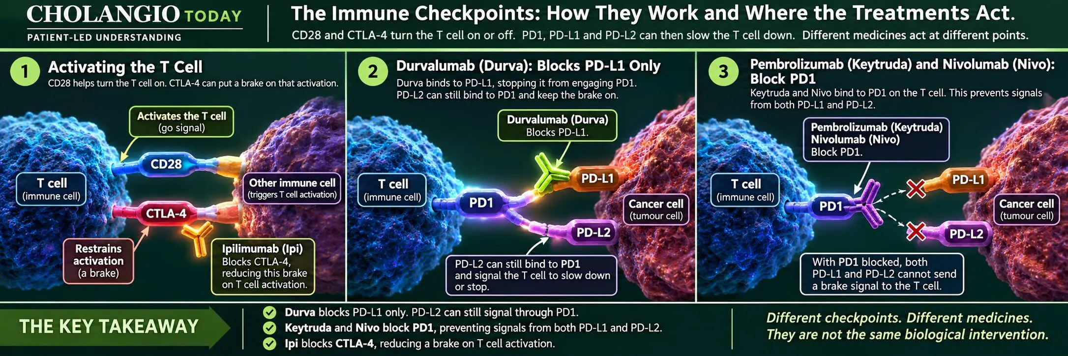
It does not mean that PD1 treatment must succeed where Durva did not.
But it means that progression after Durva cannot, by itself, establish that blocking PD1 would be biologically ineffective.
And that raises a question about the clinical trials frequently used to discuss these treatments.
TOPAZ-1 and KEYNOTE-966: What did they actually test?
TOPAZ-1 investigated durvalumab alongside gemcitabine and cisplatin in patients with previously untreated advanced biliary tract cancer.
KEYNOTE-966 investigated pembrolizumab alongside gemcitabine and cisplatin in a similar first-line treatment setting.
Both studies demonstrated an overall survival benefit from adding their respective checkpoint inhibitor to chemotherapy.
The reported median overall survival was 12.8 months in the Durva combination arm of TOPAZ-1 and 12.7 months in the Keytruda combination arm of KEYNOTE-966.
Those figures are often placed alongside one another.
I can understand why.
At first glance, the outcomes appear similar.
But what question do those numbers actually answer?
TOPAZ-1 compared gemcitabine and cisplatin with or without Durva.
KEYNOTE-966 compared gemcitabine and cisplatin with or without Keytruda.
Neither study directly compared Durva with Keytruda.
The studies also differed in important details of their treatment schedules, patient populations and trial designs.
Their median survival figures cannot establish that the two medicines are equally effective.
More importantly for this investigation, both trials studied a combination of chemotherapy and checkpoint inhibition.
They did not isolate the effects of PD1 monotherapy in patients who had exhausted conventional treatment.
Nor did they investigate nivolumab and ipilimumab as a combined PD1 and CTLA-4 approach after conventional treatment.
Those are different treatment questions.
Why the chemotherapy combination matters
Chemotherapy and immunotherapy can interact.
Chemotherapy damages cancer cells, but it can also affect immune cells and the environment in which they operate.
Depending on the medicine, dose, timing and tumour biology, those effects may support or interfere with aspects of an immune response.
For example, cancer-cell damage may release material that helps the immune system recognise the tumour.
But chemotherapy can also reduce populations of rapidly dividing immune cells.
The combined effect is not necessarily the same in every patient or every tumour.
That makes it important not to assume that a chemotherapy and PD1 combination reveals everything about the potential of PD1 blockade on its own.
We cannot conclude that chemotherapy suppressed Keytruda's effect in KEYNOTE-966.
The trial does not establish that.
But neither can we use KEYNOTE-966 to establish that Keytruda alone cannot work after chemotherapy has ended.
That was not the treatment being tested.
This is the distinction I want patients to recognise.
A trial can provide strong evidence for the treatment it investigated without answering every question about the individual medicines involved.
What about CTLA-4 and PD1 together?
There is another distinction.
Earlier in this investigation, we examined how CTLA-4 helps regulate T-cell activation, while PD1 can restrain activated T cells.
Blocking both checkpoints is not the same biological intervention as blocking PD1 alone.
And neither approach is identical to combining a PD1 inhibitor with gemcitabine and cisplatin.
The melanoma studies demonstrated responses among PD-L1-negative patients receiving nivolumab and ipilimumab together.
Biliary tract cancer studies have also documented responses to this combination.
Those findings do not establish that the combination will benefit every patient with cholangiocarcinoma.
The treatment can carry substantial immune-related risks, particularly when two checkpoints are blocked.
But it is a distinct approach supported by its own clinical observations.
Its potential should not be dismissed merely because KEYNOTE-966 did not demonstrate an obvious survival advantage over TOPAZ-1 in an informal comparison.
The studies did not test the same question.
When the patient has already exhausted conventional treatment
This is where the distinction becomes particularly important.
A patient who has exhausted conventional treatment is not asking which first-line combination should have been prescribed months or years earlier.
That decision has already been made.
The patient is asking whether another treatment opportunity remains.
They may have no established conventional treatment left that offers a reasonable prospect of benefit.
At that point, being told that KEYNOTE-966 produced results similar to TOPAZ-1 does not answer their question about PD1 monotherapy or PD1 and CTLA-4 combination treatment.
It answers a different question about first-line treatment combinations.
Nor does previous Durva progression prove that subsequent PD1 treatment cannot work.
A clinician may reasonably explain that the evidence for a particular treatment sequence is limited, that access may be difficult, or that the risks outweigh the expected benefit for an individual patient.
Those are legitimate considerations.
But they should be identified for what they are.
A lack of established evidence supporting routine use is not the same as evidence demonstrating that a treatment cannot work.
And a clinical judgement that a treatment is not justified is not the same as proving that the relevant biology has no remaining opportunity.
For the patient who has reached the end of conventional treatment, those distinctions deserve careful consideration.
TOPAZ-1 and KEYNOTE-966 answer first-line combination-treatment questions. They do not close every question about PD1 monotherapy or combined PD1 and CTLA-4 blockade.
08 | PD-L1 Negative. The Evidence That Keeps an Option Open.
The investigation began with responses in PD-L1-negative melanoma patients.
The next important finding was that responses to PD1 treatment had also been documented in advanced biliary tract cancer, including a patient classified as PD-L1 negative.
Until this point, the most striking clinical observations had come from melanoma.
They established that patients classified as PD-L1 negative could respond to PD1 inhibitors.
But my purpose was to investigate what the evidence could mean for patients with cholangiocarcinoma.
That required looking at the cancer family itself.
KEYNOTE-158: The evidence reaches biliary tract cancer
KEYNOTE-158 investigated pembrolizumab, or Keytruda, in 104 patients with advanced biliary tract cancer whose previous treatments had failed.
Biliary tract cancer includes cholangiocarcinoma, which begins in the bile ducts, and other cancers of the biliary system.
Six of the 104 patients experienced an objective response.
That represented 5.8% of the study population.
But within the overall result was a finding of particular importance to this investigation.
Thirty-four patients were classified as PD-L1 negative.
One of those 34 patients responded to Keytruda.
That represented 2.9%.
One patient among 34.
A small proportion, certainly.
But a documented clinical response despite negative PD-L1 classification.
The published subgroup results did not identify whether that particular patient had cholangiocarcinoma or another biliary tract cancer.
We cannot assign a cancer subtype that the published findings did not establish.
Nevertheless, the observation had moved beyond melanoma.
A patient with advanced biliary tract cancer had responded to PD1 treatment despite being classified as PD-L1 negative.
What does a low response rate tell us?
The overall response rate in KEYNOTE-158 was low.
That matters.
Most patients did not achieve an objective response.
But the overall percentage is not the complete story.
Some patients who responded experienced durable responses.
The published study reported responses lasting from at least 6.2 months to more than two years.
For a patient with advanced cancer, duration matters.
A treatment that produces no response in most patients may still provide a meaningful opportunity for the smaller group who respond.
That does not justify promising benefit to the next patient.
It raises a different question.
Can we understand what distinguishes the patients who respond from those who do not?
And when the treatment has already produced responses in patients classified as PD-L1 negative, should that negative classification alone be used to exclude another patient?
The observation does not support such an automatic conclusion.
KEYNOTE-158 also examined microsatellite instability.
Among patients whose MSI status was assessed, none were classified as MSI-high, including those who responded.
The recorded responses were therefore not confined to patients with that particular DNA repair-related characteristic.
This does not mean MSI status is unimportant.
It means that the observed responses cannot simply be explained away as responses occurring only in the MSI-high group.
Nivolumab: Responses in cholangiocarcinoma
The next study brought the investigation directly into cholangiocarcinoma.
In a Phase 2 study led by Kim and colleagues, researchers investigated nivolumab in patients with advanced biliary tract cancer whose previous treatments had failed.
The investigators reported responses according to where the cancer began.
Among 28 patients with intrahepatic cholangiocarcinoma, which begins in the bile ducts inside the liver, six experienced an objective response.
Among five patients with extrahepatic cholangiocarcinoma, which begins in the bile ducts outside the liver, two experienced an objective response.
Across 46 patients evaluable for response, investigators reported 10 responses, representing 22%.
An independent central review confirmed five responses, representing 11%.
Those assessments produced different response figures, and that distinction matters.
But both documented patients responding to nivolumab.
The study also examined PD-L1 expression.
Most responders had tumours classified as PD-L1 positive under its testing method.
It therefore did not establish a reliable response rate specifically for PD-L1-negative cholangiocarcinoma.
What it demonstrated was different.
Nivolumab had produced measurable responses in patients with cholangiocarcinoma.
What about blocking two immune checkpoints?
Researchers also investigated whether blocking two different immune checkpoints could produce responses in biliary tract cancers.
Nivolumab blocks PD1.
Ipilimumab blocks CTLA-4, which regulates a different part of T-cell activation.
Together, the medicines release two distinct immune restraints.
The CA209-538 study documented responses in advanced biliary tract cancer.
The Australian MoST-CIRCUIT trial subsequently investigated the same combination in patients with intrahepatic cholangiocarcinoma and gallbladder cancer.
These studies added further evidence that checkpoint treatments could produce responses in biliary tract cancers.
Because nivolumab and ipilimumab were administered together, the results could not establish how much each medicine contributed to individual responses.
Nor do these findings establish that the combination is suitable for every patient.
But they do establish that this is a treatment approach with its own clinical evidence.
It should be assessed on that evidence rather than dismissed through the results of a different treatment combination.
What have we established?
The studies now provided several distinct observations.
In melanoma, patients classified as PD-L1 negative had responded to PD1 inhibitors.
In KEYNOTE-158, a patient with advanced biliary tract cancer had responded to Keytruda despite negative PD-L1 classification.
In the study led by Kim and colleagues, patients with cholangiocarcinoma had responded to nivolumab.
Other biliary tract cancer studies documented responses to combinations involving PD1 and CTLA-4 blockade.
These findings do not tell us which individual patient will respond next.
Nor do they establish that every patient should receive the same treatment.
But the evidence does establish that PD1 treatment can produce responses in these clinical settings, including responses despite negative PD-L1 classification.
The question for the patient is no longer whether such responses have ever occurred.
They have.
The question is what should happen when this evidence is brought into a consultation by a patient who may have exhausted every conventional treatment option.
We do not inflate the few into the many. But neither do we allow the few to disappear because they are few.
09 | When the Evidence Reaches the Patient
A negative PD-L1 result and previous Durva progression are clinical facts to consider.
Neither, by itself, establishes that PD1 blockade cannot work.
And neither should automatically end the discussion when a patient asks whether another treatment opportunity remains.
I write this investigation as a survivor of Stage IV cholangiocarcinoma and as someone who has spent years working directly with patients and their families.
Through the Cholangiocarcinoma Foundation Australia, which Claire and I founded, I have worked directly with patients and their families over many years, across Australia and multiple countries internationally, helping them understand their disease, examine their medical reports, identify treatment opportunities, respond to progression and navigate decisions when conventional treatments have been exhausted.
This experience has given me a perspective across the full course of cholangiocarcinoma, from diagnosis and treatment through to progression, survival and the opportunities that can be lost along the way.
I am not a medical practitioner or laboratory scientist. My expertise has been built through lived experience, years of working directly with patients, investigating disease biology and clinical evidence, and developing practical systems that turn understanding into action.
The foundation Claire and I founded is solutions-based. We do not direct our resources towards awareness or advocacy campaigns. Our work concentrates on identifying problems, understanding their causes and delivering practical responses that can make a difference to patients.
My investigations and writing through Cholangio Today are my own independent initiative.
I undertake this work because I believe experience carries a responsibility to investigate what may help the next patient.
The findings are intended to benefit the wider cholangiocarcinoma community, including the foundation we founded, the patients and families it serves, and the many supporters, medical professionals, scientists, researchers and pharmaceutical organisations whose work contributes to improving patient outcomes.
I value those contributions. They make much of what is possible today possible.
But this investigation, the questions it pursues and the conclusions I draw are my own.
I begin with a documented clinical observation, examine what the evidence establishes, identify what remains unanswered and bring those findings back to the decisions patients face.
That is experience put to work.
What happens when conventional treatment has ended?
Imagine a patient with advanced cholangiocarcinoma.
They have received gemcitabine, cisplatin and Durva.
Their cancer has progressed.
They have undergone further treatment, perhaps chemotherapy, targeted therapy or another available approach.
Eventually, their oncologist explains that there are no further conventional treatment options to recommend.
For the patient and their family, this is not an abstract discussion about clinical trial design.
It is a question about survival.
What else can be considered?
The patient learns that Keytruda and Nivo block PD1 rather than PD-L1.
They discover that responses to PD1 inhibitors have been documented in patients classified as PD-L1 negative.
They learn that responses have also occurred in advanced biliary tract cancer.
They may also learn that blocking PD1 together with CTLA-4 represents a different biological approach.
They take that evidence to their oncologist.
What should happen next?
The difference between insufficient evidence and impossibility
There may not be enough evidence to recommend PD1 treatment routinely after Durva progression in biomarker-unselected cholangiocarcinoma.
That is a statement about the available clinical evidence.
It is not proof that PD1 treatment cannot work in an individual patient.
The distinction matters because patients frequently encounter explanations that compress these different meanings.
"You have already tried immunotherapy."
"Your PD-L1 test was negative."
"KEYNOTE-966 did not show Keytruda was better than Durva."
"There is no evidence that it works after Durva."
Each statement needs to be examined carefully.
A negative PD-L1 classification does not mean the patient has no PD1 pathway capable of being inhibited.
Previous Durva treatment does not mean the patient has received a medicine that blocks both PD1 ligands.
KEYNOTE-966 did not compare PD1 monotherapy or PD1 and CTLA-4 combination treatment after conventional therapies had been exhausted.
And an absence of direct evidence supporting a particular treatment sequence is not proof that the treatment is biologically incapable of working.
None of these distinctions guarantees benefit.
But they challenge the reasoning that would turn uncertainty into automatic exclusion.
What should the oncologist consider?
The treating oncologist has responsibilities that extend beyond this investigation.
The patient's current health, liver function, previous treatments, other medical conditions and risk of serious side effects all matter.
So do genomic findings, mismatch repair status, microsatellite instability, tumour mutational burden and other relevant clinical information.
A patient may have another treatment or clinical trial supported by stronger evidence.
PD1 treatment may be inappropriate because of the patient's medical condition or the risks involved.
Combining PD1 and CTLA-4 blockade can increase the risk of serious immune-related complications.
And when the expected chance of benefit is small or uncertain, that must be explained.
These are legitimate considerations.
The patient deserves to understand them.
But if a treatment is declined, I want the reason to be clear.
Is it because the patient is medically unsuitable?
Is there a better treatment opportunity?
Is the risk judged to outweigh the possible benefit?
Is access unavailable?
Or is the decision based on the assumption that previous Durva treatment, a negative PD-L1 result or the results of KEYNOTE-966 have already answered every relevant question?
Those are not equivalent reasons.
What patient-led investigation means
Our Patient-Led Declaration places the patient's understanding and survival opportunity at the centre of the work.
That responsibility requires more than repeating established treatment pathways.
It requires examining what the evidence demonstrates, identifying what remains unanswered, and asking whether an overlooked observation could affect what happens next for a patient.
This is not an attempt to practise medicine.
It is not a request to ignore established guidelines or the professional responsibilities of the treating team.
It is an insistence that the patient should not lose a potential opportunity because a question was never properly examined.
I am not asking the oncologist to guarantee a response.
I am asking for the clinical evidence to be considered in the circumstances of the individual patient.
If the answer is no, I want the patient to understand why.
And if another experienced specialist might identify an opportunity, I want that question raised while the patient remains able to act.
The question I would ask
If I were sitting with a patient who had exhausted conventional treatment, I would encourage them to ask their oncologist:
"Given that patients classified as PD-L1 negative have responded to PD1 inhibitors, and given that Durva and PD1 inhibitors do not act on identical molecular targets, is there a reasonable clinical basis to consider Keytruda, Nivo or a PD1 and CTLA-4 combination in my circumstances?"
I would then ask:
"If you do not recommend it, what specific evidence, medical risk or better treatment opportunity supports that decision?"
Those are reasonable questions.
They do not require the patient to claim that the treatment will work.
They require the treating team to explain whether the evidence supports an opportunity, whether the patient is suitable, and what can reasonably be done next.
The patient does not need to prove that a treatment will work before asking for the evidence to be considered.
10 | The Evidence Matters Most While You Can Still Act
Understanding a treatment opportunity matters most while the patient remains able to act on it.
For some patients, that means planning ahead while established treatment is still working.
For others, it means facing a much harder moment.
Conventional treatments have been exhausted.
The cancer continues to progress.
The patient and their family have been told that no further standard treatment is available.
Yet the patient may still be medically fit enough to consider another approach.
This is often when the question about Keytruda, Nivo or combined checkpoint treatment becomes urgent.
Not because the patient expects a miracle.
But because they want to know whether every reasonable opportunity has been considered before the door closes.
Why time changes the decision
Advanced cholangiocarcinoma can progress quickly.
A patient who is strong enough to consider another treatment today may not remain well enough indefinitely.
Declining liver function, infections, complications and loss of physical strength can make further treatment increasingly difficult.
This means that time is not simply a date on a treatment calendar.
Time can determine whether another treatment remains possible.
Imagine an open doorway.
The doorway does not promise what will happen on the other side.
But if the patient becomes too unwell to enter, the opportunity may disappear regardless of what treatment might have achieved.
That is why I believe these discussions should occur early enough for the patient to act on the decision.
It does not mean abandoning a treatment that is currently working.
It means understanding the next reasonable options before the current treatment stops working.
And if conventional treatments have already ended, it means asking for an informed assessment without unnecessary delay.
What should happen next?
If a patient and their treating team decide that PD1 treatment may offer a reasonable opportunity, the next question is access.
A medicine cannot help a patient who cannot obtain it.
An appropriate clinical trial may provide a pathway.
Depending on the country, clinical circumstances and applicable rules, other possibilities may include an approved biomarker-based indication, an off-label prescription, a special-access arrangement or a manufacturer-supported access programme.
In Australia, approval and funding are different questions.
A medicine may be legally prescribable without being routinely funded through the Pharmaceutical Benefits Scheme for that particular cancer or treatment setting.
The Therapeutic Goods Administration has special-access pathways for certain circumstances, but access is not automatic.
The treating doctor must determine what is clinically appropriate and which pathway, if any, is available.
Ask what documentation is required, who will make the application, whether the treatment can be funded and how long the process may take.
If a specialist second opinion is needed, establish that promptly.
An opportunity can be lost not only because a clinical decision was delayed, but because the steps needed to implement it began too late.
Five points to take into the consultation
You do not need to remember every trial, every biological mechanism or every response percentage.
Carry these five points into the conversation.
First. PD-L1 negative does not mean PD1 cannot work.
Clinical trials have documented responses in patients classified as PD-L1 negative.
Second. Durva is not the same molecular treatment as Keytruda or Nivo.
Durva blocks PD-L1. Keytruda and Nivo block PD1, preventing inhibitory signals through that receptor from both PD-L1 and PD-L2.
Previous Durva treatment alone does not prove that PD1 treatment cannot work.
Third. TOPAZ-1 and KEYNOTE-966 do not answer every checkpoint-treatment question.
They investigated first-line chemotherapy and checkpoint combinations.
They did not establish that PD1 monotherapy or combined PD1 and CTLA-4 treatment cannot work after conventional treatment has been exhausted.
Fourth. There is evidence in biliary tract cancer.
KEYNOTE-158 documented a PD-L1-negative response, and other studies documented PD1 treatment responses in cholangiocarcinoma.
Combination studies have also documented responses involving PD1 and CTLA-4 blockade.
Fifth. I am requesting that the evidence be applied to my individual circumstances.
This investigation has established that PD1 inhibitors have produced responses in patients classified as PD-L1 negative, including a documented response in advanced biliary tract cancer.
I am asking you to assess access to Keytruda, Nivo or an appropriate PD1 and CTLA-4 combination, rather than exclude these treatments solely because my PD-L1 result was negative or my cancer progressed after gemcitabine, cisplatin and Durva.
If another clinical factor makes treatment unsuitable, please explain it. If treatment is clinically reasonable, please help me investigate the available access pathways.
What this investigation has revealed
I began with a clinical observation that caught my attention.
In CheckMate 067, 41.3% of patients classified as PD-L1 negative responded to nivolumab alone.
That observation led me into the discoveries of Allison and Honjo, the biology of immune checkpoints, the limitations of tumour biopsies and the changing relationship between cancer and the immune system.
It raised questions about what PD-L1 testing measures, what PD-L2 may contribute and whether the immune system's earlier activity can remain invisible in a later biopsy.
The investigation then reached biliary tract cancer and cholangiocarcinoma, where published studies documented further responses to PD1 treatment.
It also exposed an important distinction between clinical trials.
A study investigating chemotherapy combined with a checkpoint inhibitor does not automatically answer questions about checkpoint treatment alone or about blocking two different immune controls together.
Nor does previous treatment with a PD-L1 inhibitor establish that a PD1 inhibitor cannot work.
Some questions remain unresolved.
We do not yet know every mechanism that explains the PD-L1-negative responders, or how to identify every future responder.
We do not have reliable evidence that every patient who has progressed after Durva will benefit from subsequent PD1 treatment.
And we cannot promise that a patient who has exhausted conventional treatment will respond to another checkpoint approach.
But uncertainty about the explanation does not undo the clinical observations.
For the patient, that distinction has a consequence.
A treatment opportunity cannot be evaluated properly if the relevant question is dismissed before it is examined.
And the patient may not have the time to wait until every biological question is answered.
That is why I have pursued this investigation.
Not to replace the oncologist's judgement, but to ensure that the patient is better equipped to question, understand and participate in the decision.
Not to turn limited evidence into a promise, but to prevent uncertainty from being mistaken for proof of impossibility.
And not to claim that every patient should receive PD1 treatment, but to ask that every medically suitable patient facing this question receives an informed, individual assessment.
The Patient-Led Conclusion
A PD-L1 result is an indicator. It is not a complete account of the immune system, nor is a negative result proof that PD1 treatment cannot work.
The clinical evidence has documented responses to PD1 inhibitors in patients classified as PD-L1 negative.
For patients with advanced cholangiocarcinoma whose cancer has progressed after gemcitabine, cisplatin and Durva, that evidence deserves consideration, particularly when conventional treatment options have been exhausted.
Previous Durva treatment does not establish that blocking PD1 will be ineffective. Nor do the first-line results of TOPAZ-1 and KEYNOTE-966 answer every question about PD1 monotherapy or combined PD1 and CTLA-4 blockade.
The patient should understand their PD-L1 result, what it measures and what it cannot establish.
They should also be able to ask for an individual assessment of PD1 treatment, including its evidence, risks, alternatives and possible access pathways.
A treatment may be declined for sound clinical reasons. But a negative PD-L1 result alone should not be the reason.
PD-L1 negative does not mean PD1 cannot work. After gemcitabine, cisplatin and Durva, do not let a negative PD-L1 result alone close the door to PD1 treatment.
To My Treating Oncologist
Please read the accompanying investigation and review the clinical evidence it brings together. I am requesting an individual assessment of whether PD1 treatment could provide another treatment opportunity in my circumstances.
I understand that a response cannot be guaranteed. I also understand that pembrolizumab (Keytruda), nivolumab (Nivo) and combinations involving ipilimumab (Ipi) carry risks, including potentially serious immune-related complications.
However, clinical trials have documented responses to PD1 inhibitors in patients classified as PD-L1 negative.
KEYNOTE-158 documented a PD-L1-negative response in advanced biliary tract cancer. Other studies have demonstrated responses to nivolumab in cholangiocarcinoma and to combined PD1 and CTLA-4 blockade in biliary tract cancers.
I recognise that these findings do not establish how likely I am to respond, particularly if my cancer has progressed after gemcitabine, cisplatin and durvalumab (Durva).
Nevertheless, Durva blocks PD-L1, whereas Keytruda and Nivo block PD1, preventing inhibitory signalling through that receptor from both PD-L1 and PD-L2.
I also understand that TOPAZ-1 and KEYNOTE-966 investigated first-line chemotherapy and checkpoint inhibitor combinations. They did not establish whether PD1 monotherapy or combined PD1 and CTLA-4 treatment would benefit a patient in my circumstances after conventional treatment has been exhausted.
I am asking that neither my negative PD-L1 result nor previous Durva treatment be used alone to exclude consideration of these treatments.
Please assess the evidence alongside my current medical condition, liver function, previous treatments, molecular testing, remaining treatment options and the risks involved.
If treatment is clinically reasonable, please help me investigate an appropriate access pathway, including clinical trials or other available arrangements.
If you believe treatment should not proceed, please explain the specific reasons, including whether another treatment offers a better opportunity or whether the risks outweigh the potential benefit.
I am not asking you to guarantee a response.
I am asking for the evidence to be considered, the decision to be explained and any reasonable opportunity to be assessed while I am still able to act.
Five Points to Take to Your Appointment
You do not need to remember every trial or understand every biological explanation. Carry these five points into the conversation.
- PD-L1 negative does not mean PD1 cannot work. Clinical trials have documented responses in patients classified as PD-L1 negative.
- Durva is not the same molecular treatment as Keytruda or Nivo. Durva blocks PD-L1. Keytruda and Nivo block PD1. Previous Durva treatment alone does not prove that PD1 blockade cannot work.
- There is evidence in biliary tract cancer. KEYNOTE-158 documented a PD-L1-negative response. Other studies documented responses to PD1 treatment in cholangiocarcinoma and to combined PD1 and CTLA-4 blockade in biliary tract cancers.
- KEYNOTE-966 does not close this question. It investigated Keytruda alongside first-line chemotherapy. It did not establish that Keytruda or Nivo alone, or Nivo combined with Ipi, cannot work after conventional treatment has been exhausted.
- I am requesting an individual treatment assessment. Please assess whether these treatments are clinically reasonable in my circumstances and, if appropriate, help me pursue access. If the answer is no, what specific evidence, medical risk or better treatment option supports that decision? Should I seek a specialist second opinion?
I do not need a guarantee. I need the evidence considered, the decision explained and the opportunity preserved wherever reasonably possible.
References & Source Material
The clinical results, biological mechanisms and patient-led questions examined in this investigation are grounded in original clinical trials, foundational checkpoint research, cholangiocarcinoma biology, drug-mechanism evidence and regulatory guidance.
What Happened to the Patients?
These sources support the response, complete-response and durability evidence that first exposes the PD-L1-negative responder question.
- Larkin J, Chiarion-Sileni V, Gonzalez R, et al. Combined Nivolumab and Ipilimumab or Monotherapy in Untreated Melanoma. New England Journal of Medicine. 2015;373:23-34. CheckMate 067. Includes the 41.3% objective response rate among patients classified as PD-L1 negative who received nivolumab, together with the broader nivolumab, nivolumab plus ipilimumab and ipilimumab treatment-arm results. DOI: 10.1056/NEJMoa1504030
- Robert C, Long GV, Brady B, et al. Nivolumab in Previously Untreated Melanoma without BRAF Mutation. New England Journal of Medicine. 2015;372:320-330. CheckMate 066. Reports an objective response rate of 33.1% among nivolumab-treated patients with negative or indeterminate PD-L1 status. DOI: 10.1056/NEJMoa1412082
- Robert C, Schachter J, Long GV, et al. Pembrolizumab versus Ipilimumab in Advanced Melanoma. New England Journal of Medicine. 2015;372:2521-2532. KEYNOTE-006. Establishes the original phase 3 pembrolizumab versus ipilimumab treatment results used as context in this investigation. DOI: 10.1056/NEJMoa1503093
- Schachter J, Ribas A, Long GV, et al. Pembrolizumab versus Ipilimumab for Advanced Melanoma: Final Overall Survival Results of a Multicentre, Randomised, Open-Label Phase 3 Study (KEYNOTE-006). The Lancet. 2017;390(10105):1853-1862. Longer follow-up from KEYNOTE-006, supporting the continuing assessment of response and survival after pembrolizumab. DOI: 10.1016/S0140-6736(17)31601-X
- Robert C, Ribas A, Schachter J, et al. Pembrolizumab versus Ipilimumab in Advanced Melanoma (KEYNOTE-006): Post-hoc 5-Year Results. Lancet Oncology. 2019;20(9):1239-1251. Five-year follow-up, demonstrating why duration of follow-up must be distinguished from response status recorded at an earlier assessment. DOI: 10.1016/S1470-2045(19)30388-2
- Postow MA, Chesney J, Pavlick AC, et al. Nivolumab and Ipilimumab versus Ipilimumab in Untreated Melanoma. New England Journal of Medicine. 2015;372:2006-2017. CheckMate 069. Supports the nivolumab plus ipilimumab response evidence, including responses among patients classified as PD-L1 negative. DOI: 10.1056/NEJMoa1414428
- Long GV, Atkinson V, Cebon JS, et al. Standard-Dose Pembrolizumab Plus Alternate-Dose Ipilimumab in Advanced Melanoma: KEYNOTE-029 Cohort 1C, Long-Term Results. Clinical Cancer Research. Among the reported PD-L1-negative subgroup, 50.0% achieved an objective response and 33.3% achieved a complete response. The broader cohort also provides longer-term response-duration context. DOI: 10.1158/1078-0432.CCR-20-0177
- Eisenhauer EA, Therasse P, Bogaerts J, et al. New Response Evaluation Criteria in Solid Tumours: Revised RECIST Guideline (Version 1.1). European Journal of Cancer. 2009;45(2):228-247. RECIST 1.1. Provides the standard response definitions underlying partial response, complete response and progressive disease. DOI: 10.1016/j.ejca.2008.10.026
What Biology Could Explain the Observation?
These sources establish adaptive and dynamic PD-L1 biology, on-treatment immune change, PD-L2 biology and the molecular distinction between blocking PD1 and blocking PD-L1.
- Taube JM, Anders RA, Young GD, et al. Colocalization of Inflammatory Response with B7-H1 Expression in Human Melanocytic Lesions Supports an Adaptive Resistance Mechanism of Immune Escape. Science Translational Medicine. 2012;4(127):127ra37. Evidence linking tumour immune activity, interferon-gamma signalling and adaptive PD-L1 expression. DOI: 10.1126/scitranslmed.3003689
- Spranger S, Spaapen RM, Zha Y, et al. Up-Regulation of PD-L1, IDO, and Tregs in the Melanoma Tumor Microenvironment Is Driven by CD8+ T Cells. Science Translational Medicine. 2013;5(200):200ra116. Supports the relationship between active tumour-reactive CD8+ T cells and adaptive changes in the tumour immune environment, including PD-L1. DOI: 10.1126/scitranslmed.3006504
- Vilain RE, Menzies AM, Wilmott JS, et al. Dynamic Changes in PD-L1 Expression and Immune Infiltrates Early During Treatment Predict Response to PD1 Blockade in Melanoma. Clinical Cancer Research. 2017;23(17):5024-5033. Paired biopsies before and early during pembrolizumab or nivolumab treatment demonstrated dynamic changes in PD-L1 and immune-cell infiltration. Early on-treatment biology provided information not captured by the pretreatment PD-L1 measurement alone. DOI: 10.1158/1078-0432.CCR-16-0698
- Yearley JH, Gibson C, Yu N, et al. PD-L2 Expression in Human Tumors: Relevance to Anti-PD1 Therapy in Cancer. Clinical Cancer Research. 2017;23(12):3158-3167. Establishes PD-L2 as another ligand of PD1. PD-L2 was detected without PD-L1 in subsets of tumours and was associated with pembrolizumab response independently of PD-L1 in the studied head-and-neck cancer population. DOI: 10.1158/1078-0432.CCR-16-1761
- Current nivolumab prescribing information. Nivolumab binds PD1 and blocks its interaction with both PD-L1 and PD-L2. This establishes the molecular distinction between PD1 blockade and PD-L1 blockade. Nivolumab prescribing information
- Current pembrolizumab prescribing information. Pembrolizumab binds PD1 and blocks its interaction with PD-L1 and PD-L2. Pembrolizumab prescribing information
- Current durvalumab prescribing information. Durvalumab binds PD-L1 and blocks PD-L1 interactions with PD1 and CD80. The prescribing information also recognises that PD-L1 expression can be induced by inflammatory signals including interferon-gamma. Because durvalumab targets PD-L1 rather than PD1, it does not block PD-L2 from binding PD1. Durvalumab prescribing information
- Leach DR, Krummel MF, Allison JP. Enhancement of Antitumor Immunity by CTLA-4 Blockade. Science. 1996;271(5256):1734-1736. Foundational experimental evidence showing that blockade of CTLA4 could enhance anti-tumour immune responses. DOI: 10.1126/science.271.5256.1734
- Ishida Y, Agata Y, Shibahara K, Honjo T. Induced Expression of PD1, a Novel Member of the Immunoglobulin Gene Superfamily, upon Programmed Cell Death. EMBO Journal. 1992;11(11):3887-3895. The original report from Tasuku Honjo's laboratory describing PD1. DOI: 10.1002/j.1460-2075.1992.tb05481.x
- The Nobel Assembly at Karolinska Institutet. The 2018 Nobel Prize in Physiology or Medicine: Discovery of Cancer Therapy by Inhibition of Negative Immune Regulation. Scientific background to the Nobel Prize awarded jointly to James P. Allison and Tasuku Honjo for their independent checkpoint discoveries. Nobel Prize scientific background
What Does the Evidence Show in Cholangiocarcinoma?
These sources bring the investigation into cholangiocarcinoma: the immune-cold tumour environment, inducible PD-L1 biology, limitations of baseline PD-L1 measurement, treatment-induced immune change, checkpoint treatment outcomes, biomarkers and treatment access.
- Loeuillard E, Conboy CB, Gores GJ, Ilyas SI. Immunobiology of Cholangiocarcinoma. JHEP Reports. 2019;1(4):297-311. Describes immune-hot and immune-cold cholangiocarcinoma. T-cell-infiltrated or immune-hot CCA is characterised by greater CD8+ T-cell infiltration, interferon-gamma activity and increased checkpoint molecules including PD-1 and PD-L1. Immune-cold CCA lacks this T-cell-rich environment and contains prominent immunosuppressive components. DOI: 10.1016/j.jhepr.2019.06.003
- Gani F, Nagarajan N, Kim Y, et al. Programmed Death Ligand 1 Expression in Human Intrahepatic Cholangiocarcinoma and Its Association with Prognosis and CD8+ T-Cell Immune Responses. Human intrahepatic cholangiocarcinoma tissue and experimental work examining the relationship between PD-L1 and CD8+ T-cell responses. Human iCCA cell lines exposed to interferon-gamma showed increased PD-L1 expression, supporting the investigation into inducible PD-L1 biology. PubMed: 30323667
- Mody K, Starr J, Saul M, et al. Patterns and Genomic Correlates of PD-L1 Expression in Patients with Biliary Tract Cancers. Journal of Gastrointestinal Oncology. 2019;10(6):1099-1109. Examined 652 biliary tract cancers using a defined PD-L1 assay and threshold. PD-L1 positivity was 8.6% overall, including 7.3% in intrahepatic cholangiocarcinoma and 5.2% in extrahepatic cholangiocarcinoma. These percentages belong to that assay and threshold. DOI: 10.21037/jgo.2019.08.08
- Phase II pembrolizumab plus CAPOX study in advanced biliary tract carcinoma. Included paired tumour biopsies, PD-L1 and immune-infiltrate assessment, together with transcriptomic and genomic analyses. Demonstrated the feasibility of examining how biliary tract cancer immune biology changes during treatment. PubMed: 35274717
- Phase Ib paired-biopsy study of anti-TIM-3 plus anti-PD-1 treatment in advanced biliary tract cancer. Exploratory paired tumour biopsies showed increased intratumoral CD8 T-cell density and upregulation of gene signatures related to interferon-gamma signalling, antigen presentation and T-cell activation. These changes were not shown to correlate clearly with efficacy. PubMed: 41825935
- Kim RD, Chung V, Alese OB, et al. A Phase 2 Multi-institutional Study of Nivolumab for Patients With Advanced Refractory Biliary Tract Cancer. JAMA Oncology. 2020;6(6):888-894. Prospective phase 2 evidence of PD-1 blockade with nivolumab in advanced refractory biliary tract cancer. Supports the clinical response evidence discussed in Section 08. DOI: 10.1001/jamaoncol.2020.0930
- Klein O, Kee D, Nagrial A, et al. Evaluation of Combination Nivolumab and Ipilimumab Immunotherapy in Patients With Advanced Biliary Tract Cancers: Subgroup Analysis of the CA209-538 Trial. JAMA Oncology. 2020. Phase 2 evidence examining combined PD-1 and CTLA-4 blockade in advanced biliary tract cancers, including intrahepatic cholangiocarcinoma and gallbladder cancer. DOI: 10.1001/jamaoncol.2020.2814
- Oh D-Y, He AR, Qin S, et al. Durvalumab plus Gemcitabine and Cisplatin in Advanced Biliary Tract Cancer. NEJM Evidence. 2022;1(8). TOPAZ-1. Phase 3 evidence establishing the clinical benefit of adding the PD-L1 inhibitor durvalumab to gemcitabine and cisplatin in previously untreated advanced biliary tract cancer. DOI: 10.1056/EVIDoa2200015
- Kelley RK, Ueno M, Yoo C, et al. Pembrolizumab in Combination with Gemcitabine and Cisplatin Compared with Gemcitabine and Cisplatin Alone for Patients with Advanced Biliary Tract Cancer (KEYNOTE-966). The Lancet. 2023. Phase 3 evidence establishing clinical benefit from adding the PD-1 inhibitor pembrolizumab to gemcitabine and cisplatin in previously untreated unresectable or metastatic biliary tract cancer. TOPAZ-1 and KEYNOTE-966 are not head-to-head comparisons. PubMed: 37075781
- Tanaka S, Umemoto K, Kubo S, et al. Nivolumab for Treating Patients with Occupational Cholangiocarcinoma. Journal of Hepato-Biliary-Pancreatic Sciences. 2022;29:1153-1155. Documents complete responses to nivolumab in recurrent occupational cholangiocarcinoma. These observations establish that such responses can occur, but do not establish reproducible benefit across cholangiocarcinoma. DOI: 10.1002/jhbp.1215
- Nagrial A, et al. Nivolumab and Ipilimumab Combination Treatment in Patients with Advanced Intrahepatic Cholangiocarcinoma and Gallbladder Cancer: Results from the Phase II MoST-CIRCUIT Trial. Clinical Cancer Research. 2026;32(15):3203-3212. Contemporary phase 2 evidence of combined PD-1 and CTLA-4 blockade in 60 patients with advanced iCCA or gallbladder cancer. The overall objective response rate was 12%, including a 2% complete-response rate. Thirteen patients had previously received durvalumab. The study does not establish that switching from PD-L1 to PD-1 blockade improves outcomes after progression. DOI: 10.1158/1078-0432.CCR-25-4009
- Piha-Paul SA, Oh D-Y, Ueno M, et al. Efficacy and Safety of Pembrolizumab for the Treatment of Advanced Biliary Cancer: Results from the KEYNOTE-158 and KEYNOTE-028 Studies. International Journal of Cancer. 2020;147(8):2190-2198. Direct evidence for Section 08. KEYNOTE-158 reported an objective response rate of 5.8% (6/104), including one response among 34 patients classified as PD-L1 non-expressers (2.9%). KEYNOTE-028 reported an objective response rate of 13.0% (3/23). Neither study established efficacy after durvalumab progression. DOI: 10.1002/ijc.33013
- U.S. Food and Drug Administration. FDA Approves Pembrolizumab for Adults and Children with Tumour Mutational Burden-High Solid Tumours. 2020. Supports the TMB-high biomarker discussion. The U.S. tissue-agnostic indication specifies TMB of at least 10 mutations per megabase using an FDA-authorised test in qualifying previously treated unresectable or metastatic solid tumours without satisfactory alternatives. U.S. approval does not establish Australian eligibility or reimbursement. FDA: TMB-high approval
- U.S. National Library of Medicine, DailyMed. KEYTRUDA (Pembrolizumab): Current U.S. Prescribing Information. Supports the MSI-H/dMMR and TMB-high biomarker discussion. Lists the relevant U.S. tissue-agnostic pembrolizumab indications and their eligibility conditions. These indications are jurisdiction-specific and do not establish benefit for every patient with cholangiocarcinoma. Pembrolizumab prescribing information
- Australian Therapeutic Goods Administration. Special Access Scheme (SAS): Guidance for Health Practitioners Accessing Unapproved Therapeutic Goods. Supports the Australian access-pathway discussion. Off-label prescribing of an ARTG-included medicine generally does not require TGA Special Access Scheme approval. SAS primarily concerns access to unapproved therapeutic goods. Clinical appropriateness, prescribing requirements, informed consent and funding remain separate considerations. TGA: Special Access Scheme guidance
- Shroff RT, Kennedy EB, Alarcon Rozas AE, et al. Treatment of Advanced Biliary Tract Cancer: ASCO Living Guideline, Version 2026.1.0. Journal of Clinical Oncology. Published 14 October 2026. Current ASCO guidance recommends broad-based tissue DNA and RNA next-generation sequencing at diagnosis to identify actionable biomarkers. Pembrolizumab monotherapy is an option for qualifying dMMR/MSI-H advanced biliary tract cancer. For patients progressing after first-line therapy without an identified actionable biomarker, recommendations include FOLFOX with active symptom control or active symptom control alone, with further options depending on patient suitability. The guideline does not establish routine PD-1 treatment following durvalumab progression in biomarker-unselected cholangiocarcinoma. DOI: 10.1200/JCO-26-02462
What is known must remain known. What is missing must remain visible.
The evidence establishes that cholangiocarcinoma can be immune-cold, that interferon-gamma can induce PD-L1 in cholangiocarcinoma cells, that checkpoint-based treatment can alter the tumour immune environment, and that PD1 and PD-L1 inhibitors do not block identical checkpoint biology.
What the evidence does not yet establish is how much dynamic PD-L1 induction explains PD-L1-negative response in cholangiocarcinoma, how much PD-L2 contributes, or whether switching from PD-L1 blockade to PD1 blockade creates a clinically meaningful opportunity for an individual patient.
We do not inflate the few into the many. But neither do we allow the few to disappear because they are few.