Listen to the Complete Investigation
When Evidence Is Left Behind
Written and narrated by Steve Holmes.

Listen continuously or select any chapter. Your listening position is saved on this device.
My Patient Tools
Save what matters. Prepare your questions. Return to the evidence.
Send my saved information
Prepare an email containing your saved Anchors or Consultation notes. Choose a recipient and press Send in your own email application.
Highlight a passage and select Save to My Anchors. Use saved passages to prepare your consultation.
My Anchors
My Consultation
Select the evidence you want to discuss with your doctor.
Beta: Your information is saved in this browser. Export important notes. Avoid storing private information on shared devices.
Initialising Patient Tools...
01 | The Observation
PD-L1 Negative. Still Responds. What Are We Missing?
In 2019 I was in Salt Lake City, sitting among several hundred clinicians, researchers and industry representatives. Medical oncologists were presenting findings associated with KEYNOTE-028, KEYNOTE-029 and KEYNOTE-158, among the most recent immunotherapy findings available at that time.
Both KEYNOTE-028 and KEYNOTE-158 had reported a single complete response in their respective biliary tract cancer study groups. The complete response recorded in KEYNOTE-158 in 2017 was mine.
I remember the scale of that gathering, but even more clearly I remember the question I carried into rooms like that: what happens to the patient when the experts say, “We just don’t know”?
I had been questioning cancer treatment explanations for some time. The evidence I encountered did not always fit the conclusions patients were being asked to accept. There was nothing wrong with admitting uncertainty. The problem was what uncertainty could become by the time it reached a patient: a reason not to ask, not to investigate, not to consider a remaining option.
The scientists could continue studying the question. The patient had to live through the interval. In cholangiocarcinoma, that interval can determine whether a person remains well enough to receive treatment at all.
I wanted to know what had already been observed, not only what the next trial might eventually establish.
One finding in particular caught my attention.
In CheckMate 067, a clinical trial involving patients with advanced melanoma, 41.3% of patients whose tumours were classified as PD-L1 negative responded to nivolumab alone, a medicine that blocks the PD1 immune checkpoint.
The trial used a threshold of 5% tumour-cell PD-L1 staining to distinguish positive from negative results. Patients below that threshold were classified as negative.
Yet approximately four in ten of those patients experienced measurable tumour shrinkage meeting the trial's criteria for an objective response.
That was the observation.
What the result did not explain was why those patients responded.
I came to this question as a patient, not a scientist. Having experienced Stage IV cholangiocarcinoma and a complete response to immunotherapy, I wanted to understand what had made my response possible. More importantly, I wanted to understand what the science could reveal for patients who were still searching for a treatment opportunity.
The CheckMate 067 finding came from melanoma, not cholangiocarcinoma. Its response rate could not simply be transferred from one cancer to another.
But the observation itself deserved attention.
Patients had been classified as negative for a protein involved in suppressing immune activity. Yet some had responded to a treatment designed to interrupt that suppression.
How could that happen?
Inside the tumour
To understand the question, we need to look briefly inside the tumour.
Visualise a tumour as a growing collection of cells. Each cell is its own tiny Cell City, carrying the instructions and operating systems that determine how it functions.
Around those cells are blood vessels, supporting tissue and immune cells, all interacting within the same environment.
Among the immune cells are T cells. Some can recognise and attack cancer cells.
But the immune system also needs controls to prevent unnecessary damage to healthy tissue. One of those controls involves a receptor called PD1, found on activated T cells and certain other immune cells.
Think of PD1 as a brake receiver.
Two proteins can connect to that receiver: PD-L1 and PD-L2. Both can deliver signals that restrain the T cell.
These proteins are part of the body's normal protective machinery, helping prevent unnecessary immune damage. Cancer cells, however, can use this same system to weaken an immune attack.
Medicines such as nivolumab, known as Nivo, and pembrolizumab, known as Keytruda, bind to PD1 on T cells. This prevents PD-L1 and PD-L2 from binding to PD1 and applying the T cell's brake.
Unlike chemotherapy, these medicines do not directly attack cancer cells. They remove one restraint on immune cells that may already be capable of attacking.
But removing a brake is not the same as creating an immune response. The T cells must still recognise the cancer and be capable of responding.
Now consider the original observation.
A tumour classified as PD-L1 negative under the trial's threshold was not necessarily a tumour with no PD-L1 anywhere. Nor did that classification measure PD-L2 or describe every interaction between the immune system and the cancer.
The test provided information. But how much of the biology did that information capture?
The patients who responded had demonstrated something important: the negative classification had not prevented their response.
Their earlier immune histories remained unknown.
That was where the investigation began.
What had the test measured, what had it left unseen, and what might explain the responses?
The question a study sets out to answer may not be the most important question its results uncover.
02 | The Breakthrough
Two unexpected discoveries revealed how releasing the immune system’s brakes could create a new opportunity to treat cancer.
For much of modern cancer treatment, the approach was to attack the cancer itself. Surgery removed it, radiation damaged it, and chemotherapy interfered with processes cancer cells needed to survive and multiply.
But another possibility was emerging. What if the immune system could recognise the cancer, yet its own protective controls were restraining the response?
Two scientists, investigating different questions, would help reveal how those controls worked.
In the United States, James Allison was studying T-cell activation. In Japan, Tasuku Honjo’s laboratory was investigating cell biology when it identified a protein called PD1. Neither investigation began with a finished plan for the cancer medicines that followed. The treatment opportunities emerged from pursuing what the research revealed.
Allison and the activation brake
To understand Allison’s work, think of T cells as members of Cell City’s security team.
A specialised immune cell, called an antigen-presenting cell, collects evidence of a possible threat and presents it to a T cell. When the T cell recognises that evidence, it receives an initial activation signal.
Another protein on the T cell, called CD28, connects to matching proteins on the antigen-presenting cell. That connection supplies an additional signal supporting full activation.
Think of the security officer recognising the threat, then receiving authorisation to respond.
But the T cell also has a safety brake called CTLA-4.
As the T cell activates, more CTLA-4 can appear on its surface. CTLA-4 binds to the same proteins on the antigen-presenting cell that CD28 needs, generally binding more strongly. By occupying those connections, it can prevent CD28 from receiving the additional activation signal.
That is what it brakes: the signals supporting T-cell activation.
This control helps prevent excessive immune activity from damaging healthy tissue. The brake is not necessarily faulty. It may be working normally at a time when a stronger anti-cancer response is needed.
Allison’s research helped establish CTLA-4’s restraining role. He then pursued a consequential question: could blocking that restraint allow an immune attack against cancer?
Experiments in mice showed that blocking CTLA-4 could allow the immune system to eliminate established tumours. The work helped lead to ipilimumab, commonly called Ipi, which blocks CTLA-4.
Ipi can therefore reduce CTLA-4’s interference with the activation signals received through CD28. It does not identify the cancer for the T cell. It removes a restraint on the response.
Honjo and the second brake
Honjo’s discovery followed a different path.
In 1992, his laboratory identified PD1 while investigating processes associated with cell death. Its later importance as an immune brake was not what the researchers had originally been looking for. Further work established a different role: PD1 helped restrain immune activity.
Return to Cell City’s security officer.
The officer has recognised a threat and begun responding. Another instruction can now arrive, telling the officer to slow down.
PD1 is a receiver for that braking instruction. As we established in Chapter 01, two proteins, PD-L1 and PD-L2, can connect to it and reduce T-cell activity.
These are normal protective controls. Cancer can use the same system to weaken an immune attack.
Research following Honjo’s discovery helped establish PD1-blocking medicines, including nivolumab, known as Nivo, and pembrolizumab, known as Keytruda. They bind to PD1, preventing PD-L1 and PD-L2 from connecting to that receptor and applying the T cell’s brake.
The distinction between the two checkpoints is useful:
CTLA-4 helps restrain the signals supporting T-cell activation. PD1 can restrain activated T cells as they respond.
Blocking either can remove something that has hindered an anti-cancer response. The immune cells must still recognise the cancer and be capable of attacking it.
What made the discoveries matter
In 2018, Allison and Honjo shared the Nobel Prize in Physiology or Medicine for discoveries that established cancer treatment through releasing restraints on the immune system.
The medicines changed cancer treatment. But another part of their story matters to this investigation.
The discoveries were unexpected. What followed was deliberate.
They and other researchers pursued the observations, tested their meaning and developed the findings into treatments. An observation had opened a possibility before its full significance was understood.
Years later, I encountered a result involving the PD1 checkpoint that left me with an unanswered question. I approached it from a different position: as a patient whose own cancer had responded to immunotherapy.
Fortuitous discovery opens the question. Persistent investigation reveals the opportunity.
03 | The Patient-Led Consequence
An unanswered explanation must not erase an observed response, especially when the distinction could affect a patient’s treatment opportunity.
I had survived Stage IV cholangiocarcinoma. In trying to understand my own response to immunotherapy, I had begun investigating evidence that might help other patients still searching for an opportunity.
The CheckMate 067 finding introduced in Chapter 01 stayed with me. Patients classified as PD-L1 negative had responded to nivolumab. The researchers had recorded those responses; I had recognised a question their results left open.
What had the negative classification told us, and what had it failed to explain?
I kept hearing, “We just don’t know.” But there were two different things to understand: the responses had been observed; their explanation remained incomplete.
If those became blurred, a patient could receive a very different message.
How an unanswered question becomes a half-truth
Clinical evidence travels. A finding moves from a research paper into conference presentations, guidelines, treatment pathways and conversations between doctors and patients.
Along the way, it must be summarised. The danger is that an important observation can disappear while the conclusion continues travelling.
A study may answer its original question while revealing another that it was never designed to resolve. That observation may fall outside the research programme, the analysis planned or the work funded. None of those circumstances makes it disappear from the evidence.
Yet a complicated finding is harder to carry forward than a simple explanation.
“We do not understand why these patients responded” can gradually become “Patients with a negative result do not respond.”
The first statement leaves a question open. The second closes it, despite the recorded responses.
This is what I mean by a half-truth. It need not begin with anyone deliberately withholding information. It can emerge when an important part of the evidence is omitted, oversimplified or treated as less relevant than the conclusion it complicates.
The patient and their doctor may then receive an account that sounds complete without knowing what has been left behind.
The patient lives with a different clock
For a researcher, an unanswered question may become another experiment or a future trial. For a patient, it may concern a decision that cannot wait for that work to finish.
I knew what that pressure meant. When conventional treatment opportunities had run out, understanding was critical. I needed to know what remained possible while I could still act.
That experience shaped the question I brought to the evidence.
What has already been observed that could matter now?
A finding does not become useful to patients simply because it exists in a journal. It must reach the people making the decision, in a form they can understand and examine.
But what if the clinician has never encountered it? What if the observation was lost in a summary before either the doctor or patient saw it?
This is why making evidence accessible matters to both. A doctor cannot discuss a finding they have not encountered. A patient cannot ask about an opportunity they do not know exists.
A scientist may have years to investigate an unanswered question. To a cholangiocarcinoma patient they do not have time.
Following the evidence as a patient
Allison and Honjo’s work had shown the importance of pursuing observations beyond the original question. I was applying that discipline from the patient’s position.
I could read a published finding, compare it with an explanation and ask where the two did not fit. I could investigate what the test measured, look for the observation in other studies and bring the unanswered questions forward.
I did not need to explain the entire mechanism before recognising that a recorded response challenged an absolute claim that response was impossible.
Nor did a melanoma finding settle the treatment question in cholangiocarcinoma. It identified something worth investigating further.
That distinction gave the work its direction.
The evidence should determine what I could say. The patient’s need gave me a reason to keep looking.
This investigation is undertaken for people who cannot be expected to assemble that understanding alone while carrying the pressure of a cancer diagnosis. Knowledge gained through one patient’s experience should become available to the next.
Their starting point should be further forward because someone carried the evidence with them.
The next test
Recognising an unanswered question was only the beginning. I now needed to examine whether the observation that caught my attention stood alone.
Had patients without a positive PD-L1 classification responded in other trials? Had the finding appeared with another medicine that blocked PD1?
Those questions would take the investigation beyond its starting point.
If the observation was repeated, I would then need to examine the test itself and the biology it was being asked to describe. If the evidence contradicted my interpretation, that belonged in the investigation too.
The responsibility was to follow the record, including what complicated it, and make its meaning usable while patients still had decisions to make.
The unanswered question must travel with the answer when a patient’s opportunity may depend on it.
04 | Confirmation
Was CheckMate 067 an isolated result?
The CheckMate 067 observation had raised a question I could not ignore. Patients with advanced melanoma whose tumours were classified as PD-L1 negative had responded to nivolumab.
But one observation, however compelling, was not enough.
If I was going to question the conclusions being drawn from a negative PD-L1 result, I needed to subject my own interpretation to the same discipline I expected of others.
Was CheckMate 067 an isolated finding, or had other researchers observed the same thing?
A clinical trial can produce an unusual result that does not survive further investigation. Differences between patients, limitations in measurement or statistical variation can all influence what is observed.
I went looking for other trials, other patients and, importantly, other medicines that blocked the PD1 immune checkpoint.
What I found did not solve the biological mystery. It changed the strength of the evidence behind my question.
The first test: CheckMate 066
CheckMate 066 was another clinical trial involving patients with advanced melanoma. It investigated nivolumab, the same PD1-blocking medicine used in CheckMate 067.
Among patients whose PD-L1 status was classified as negative or indeterminate, 33.1% experienced an objective response to nivolumab.
Approximately one in three patients in that group experienced measurable tumour shrinkage meeting the trial's response criteria.
The classification was not identical to CheckMate 067. CheckMate 066 combined negative and indeterminate results, so the percentages could not be directly compared.
But patients without a positive baseline PD-L1 classification had responded to nivolumab in another trial.
The observation had survived its first test.
I now wanted to know whether it was peculiar to nivolumab or could also be found with another medicine acting on PD1.
The second test: KEYNOTE-006
KEYNOTE-006 investigated pembrolizumab, known as Keytruda, against ipilimumab, commonly called Ipi.
Pembrolizumab and nivolumab are different medicines, but both block PD1. If patients classified as PD-L1 negative responded to pembrolizumab, the observation would extend beyond one medicine.
In the reported PD-L1-negative subgroup, 25% of patients receiving pembrolizumab experienced an objective response, compared with 13% receiving ipilimumab.
There it was again.
Patients classified as PD-L1 negative had responded to a second medicine blocking PD1.
The studies involved different patients, treatment conditions and assessment methods. Their percentages were not interchangeable.
But the finding was repeated.
It was no longer confined to one trial or one PD1-blocking medicine.
The third test: Blocking two immune brakes
Allison and Honjo's discoveries had revealed two different controls on T-cell activity: CTLA-4 and PD1.
Researchers had begun investigating what happened when medicines blocking these checkpoints were combined.
For my investigation, these studies offered another opportunity to examine responses among patients classified as PD-L1 negative.
CheckMate 067: The combination
Among patients whose tumours expressed PD-L1 below the study's 5% threshold, CheckMate 067 reported:
- 41.3% objective response with nivolumab alone.
- 54.8% with nivolumab combined with ipilimumab.
- 17.8% with ipilimumab alone.
The combination produced responses in more than half the patients in this PD-L1-negative subgroup.
That result did not establish how much each checkpoint contributed to an individual response. Blocking two different immune controls also produced substantially greater treatment toxicity, an important consideration when assessing potential benefit.
But the finding added to the evidence that a negative PD-L1 classification did not prevent a response to checkpoint treatment.
CheckMate 069: Further evidence
CheckMate 069 also investigated nivolumab combined with ipilimumab in advanced melanoma.
It documented responses among patients with PD-L1 expression below 5%, providing further evidence that the observation extended to another trial involving combined checkpoint blockade.
The precise subgroup response percentage still requires verification against the original report.
KEYNOTE-029: What longer follow-up revealed
KEYNOTE-029 investigated pembrolizumab combined with a reduced dose of ipilimumab.
Its original 2017 report included 24 patients classified as PD-L1 negative. Twelve experienced an objective response, representing 50% of that subgroup. Four had achieved a complete response.
When researchers published longer-term follow-up in 2020, the number of complete responses among those 24 patients had increased from four to eight.
By that later assessment, one-third of the subgroup had achieved a complete response, while the total number who had experienced an objective response remained twelve.
This added something important to my investigation.
The first published assessment of a clinical trial does not necessarily reveal the eventual depth of a patient's response. Some responses deepen over time.
The subgroup was small, and its results could not predict what would happen to an individual patient, particularly someone with another cancer.
But the longer follow-up revealed something that the original assessment had not yet captured.
Response is not the same as cure
I needed to distinguish what these trial results measured.
An objective response includes partial and complete responses.
Under commonly used RECIST 1.1 criteria, a partial response generally means at least a 30% reduction in the combined diameters of selected target lesions compared with baseline.
A complete response means the disappearance of detectable disease under the study's assessment criteria.
Think of a scan as a picture of what medical imaging can detect. When the cancer is no longer detectable, that is an important result. But it does not necessarily establish that every malignant cell has been eliminated.
Duration matters too. A patient whose cancer shrinks temporarily and one whose cancer remains undetectable for years may both be recorded as responders, despite having very different outcomes.
A response rate is not a cure rate.
That distinction matters when patients interpret trial evidence. It also matters when researchers report outcomes.
The trial findings showed that responses occurred. They did not establish why those patients responded, how long every response would last or what would happen in cholangiocarcinoma.
What the combined evidence revealed
I had begun with an observation in CheckMate 067 and tested whether it stood alone.
It did not.
Across multiple melanoma trials, patients classified as PD-L1 negative, or without a positive baseline PD-L1 classification, had responded to medicines blocking PD1. The observation extended across nivolumab, pembrolizumab and combinations involving CTLA-4 blockade.
The studies were not identical, and their results could not be combined into one response rate or transferred directly to cholangiocarcinoma.
But they had established something precise.
A negative baseline PD-L1 classification did not make a response to PD1 blockade impossible.
That was no longer a question of whether such responses could occur.
They had occurred.
The unanswered question was why.
The next question was hidden in the test
The investigation had moved from one unexpected observation to a pattern documented across multiple trials.
I now needed to examine the classification itself.
When a patient's tumour is described as PD-L1 negative, what does that actually mean? What was measured, where was it measured, and what might remain unseen?
The answer begins with a biopsy.
A biopsy can accurately describe the tissue examined without revealing everything happening throughout the cancer.
05 | The Biopsy Is Just One Picture
What Does a PD-L1-Negative Biopsy Actually Tell Us?
The clinical trials had established something important. Patients whose tumours were classified as PD-L1 negative had responded to medicines that blocked PD1.
The observation was no longer confined to one study, one group of patients or one medicine. But the trials had not established why those responses occurred.
I had followed the evidence far enough to recognise that the next question might not lie in the treatment results themselves. It might lie in something that had happened much earlier, when a small piece of tumour tissue was collected, examined and given a classification.
A patient receives a pathology report. Somewhere in that report may be a result that appears straightforward: PD-L1 positive or PD-L1 negative.
To the patient, those words can sound definitive. Positive suggests that something is present. Negative suggests that it is absent.
But was that what the test actually established?
I wanted to understand what the pathologist had measured, what the result meant, and whether a negative classification could describe the tissue accurately while still leaving important parts of the cancer's biology unseen.
Because if a patient was going to be told that a treatment was unlikely to help, or was not worth considering, I wanted to know precisely what evidence supported that conclusion.
What Does the Test Actually Measure?
When a doctor requests PD-L1 testing, a pathologist examines a sample of cancer tissue using a laboratory method called immunohistochemistry, or IHC.
The method uses specially designed antibodies to detect PD-L1 protein in the tissue. The laboratory processes the sample so that cells expressing the protein can be identified under a microscope.
Think of it as applying a special marker to the tissue. The marker helps reveal where PD-L1 is detectable in the sample being examined.
The pathologist can then assess the staining according to a defined scoring method.
But the test is not measuring the entire immune system. It is measuring PD-L1 expression in the tissue available for examination, using a particular laboratory method and set of rules.
And those rules matter.
One commonly used scoring method is the Tumour Proportion Score, or TPS. It measures the percentage of viable cancer cells showing the required PD-L1 staining.
Another method is the Combined Positive Score, or CPS. This includes PD-L1 staining in certain immune cells, as well as cancer cells, and relates the count to the number of viable cancer cells.
These methods do not measure exactly the same thing.
A tumour could therefore receive different numerical results depending on the scoring method used. The meaning of those results also depends on the cancer type, the test and the treatment being considered.
This was my first important discovery about the classification.
PD-L1 negative is not a complete biological description. It is a result produced by a particular measurement and threshold.
Negative Does Not Necessarily Mean Zero
I returned to CheckMate 067.
In that study, the PD-L1-negative subgroup was defined using a threshold of 5% tumour-cell PD-L1 expression.
Patients whose measured expression fell below that threshold were classified as negative for the subgroup analysis.
That distinction is fundamental.
A tumour with measurable PD-L1 expression below 5% could be placed in the same negative category as a tumour with no detectable staining in the assessed cancer cells.
Yet those findings are not identical.
One describes expression below a chosen threshold. The other describes an absence of detectable staining under the conditions of the test.
The word negative can conceal that difference.
Imagine a measurement in which the dividing line is five. A result of four is below the line, but four is not zero.
The classification may be entirely correct according to the study's rules.
The danger arises when someone interprets the classification as meaning something more absolute than the measurement supports.
In CheckMate 067, PD-L1 negative did not necessarily mean that PD-L1 was completely absent.
It meant that the assessed tumour-cell expression was below the study's defined threshold.
I could now see why understanding the actual result, rather than simply accepting the label, mattered.
But there was another limitation.
Even if the laboratory detected no PD-L1 in the particular sample, could that result tell us what was happening throughout the entire tumour?
A Photograph of One Street in Cell City
Imagine standing in a city and taking a photograph of one street.
The photograph may be clear, accurate and taken with an excellent camera.
It can show the buildings, the people and the activity occurring in that street at that moment.
But it cannot tell you everything happening in the surrounding neighbourhoods.
It cannot show what is taking place on the other side of the city.
And it cannot reveal what happened yesterday.
A tumour biopsy has a similar limitation.
The tissue examined by a pathologist comes from a particular location. It may be a small sample taken with a needle, tissue collected during a procedure or part of a larger surgical specimen.
The pathologist can examine what is present in that material.
But cancer is not necessarily uniform throughout its structure.
One region may contain different populations of cancer cells, immune cells and supporting tissue from another.
PD-L1 expression may also vary between those regions.
A biopsy taken from one part of a tumour may therefore reveal a different pattern from a sample collected elsewhere.
This does not mean the original test was wrong.
The pathologist may have measured the available tissue correctly.
The limitation is that the sample may not represent every region of the cancer.
An accurate picture of one part is not necessarily a complete picture of the whole.
For patients with cholangiocarcinoma, this distinction deserves particular attention. Tissue may be obtained from the primary tumour, a metastatic deposit or a limited biopsy collected during a diagnostic procedure.
The location, quantity and quality of the tissue can affect what the laboratory is able to assess.
A patient may therefore have a valid pathology result without having a complete account of the biological differences across their disease.
What About the Cells Surrounding the Cancer?
There was another question hidden in the measurement.
PD-L1 is not expressed only by cancer cells.
Certain immune cells and other cells in the tumour environment can also express PD-L1.
This matters because a tumour is more than a collection of malignant cells.
Around them are immune cells, blood vessels and supporting tissue. These cells communicate through chemical signals that can influence whether the immune system attacks the cancer or is held back.
Some immune cells may be trying to recognise and attack the cancer. Others may contribute to conditions that suppress an effective immune response.
The PD1 checkpoint can operate within this environment.
If a test's scoring method focuses on PD-L1 expression in cancer cells, it may not fully describe PD-L1 expression on surrounding immune cells.
That does not make the scoring method invalid.
It means the measurement has a defined purpose and a defined boundary.
I began to see how a patient could receive a negative classification without being told exactly which cells had been assessed, which cells had contributed to the score and which biological features had not been measured.
The distinction became even more important when I returned to the mechanism of PD1 treatment.
PD-L1 Is Not the Only Braking Signal
In Chapter 02, we explored the immune checkpoint discoveries that helped make PD1 treatment possible.
PD1 is a receptor found on T cells. When PD-L1 binds to PD1, it can deliver an inhibitory braking signal that reduces T-cell activity.
But PD-L1 is not the only protein capable of engaging PD1.
There is another: PD-L2.
PD-L2 can also bind to PD1 and deliver an inhibitory braking signal.
This is why the distinction between medicines that block PD1 and medicines that block PD-L1 matters.
Pembrolizumab and nivolumab bind to PD1 itself. By occupying that receptor, they prevent both PD-L1 and PD-L2 from engaging it and applying their inhibitory braking signals.
A standard PD-L1 test, however, does not measure PD-L2 expression.
Think of PD1 as a braking control on a T cell, with PD-L1 and PD-L2 representing two different ways that control can be activated.
A test that measures PD-L1 does not automatically tell us whether PD-L2 is present or contributing to the braking process.
That is a limitation of what has been measured, not evidence that the test has malfunctioned.
Could PD-L2 have contributed to responses among patients classified as PD-L1 negative in CheckMate 067?
It is a legitimate biological question.
But the trial's response figures did not establish that PD-L2 caused those responses.
Nor did they establish that PD-L2 was present in every patient who responded.
This distinction matters throughout my investigation.
A possible explanation is not a demonstrated explanation.
The evidence tells us that PD-L2 provides another route for inhibitory signalling through PD1.
Whether that mechanism explains a particular patient's response requires additional evidence.
What Does the Pathology Report Actually Tell the Patient?
By now, the simple positive-or-negative classification had become a much more detailed question.
I wanted to know what had actually been measured.
Not because every patient needs to become a pathologist, but because a result can influence how a patient understands their treatment opportunities.
If I were helping someone review a PD-L1 result, I would want the patient and their treating team to be able to answer several questions.
Which test was used?
Different laboratory assays can use different antibodies, equipment and interpretation rules.
How was the result scored?
Was it a Tumour Proportion Score, a Combined Positive Score or another method?
What was the actual result?
Was PD-L1 expression undetectable, measurable but below the relevant threshold, or reported only as positive or negative?
What threshold was applied?
A result described as negative under one threshold may not carry the same meaning under another.
Where and when was the tissue collected?
Was it from the original tumour or a metastatic site? Was it collected recently or before earlier treatment?
And finally, was the test actually performed?
That last question may seem obvious, but it is essential.
A report that does not mention PD-L1 does not establish that the tumour is PD-L1 negative.
The test may not have been requested. There may have been insufficient tissue. The result may have been inconclusive, or it may be recorded in a separate report.
Those situations are not equivalent.
Not tested is not negative.
Inconclusive is not negative.
Below a threshold does not necessarily mean zero.
And even a result of zero in the sampled tissue does not establish that the protein was absent everywhere in the cancer.
These are distinctions that patients and doctors should be able to see clearly.
What Should Happen Earlier?
This investigation had a practical consequence.
If PD-L1 testing has been performed, the patient should be able to obtain the actual result and understand its meaning before a treatment decision depends on it.
That does not mean every patient with cholangiocarcinoma must undergo PD-L1 testing, or that a negative result automatically creates an indication for PD1 treatment.
The clinical relevance of PD-L1 varies by cancer type and treatment. Decisions must consider the broader evidence, including the patient's condition, previous treatments, other biomarkers, treatment risks and available options.
But when a result is used to support a decision, its meaning should be accurately understood.
For a patient with cholangiocarcinoma, I would want the treating team to explain whether the PD-L1 result is relevant to the specific treatment being considered, whether other biomarkers such as MSI or MMR have been assessed where appropriate, and whether specialist review could clarify an unresolved question.
I would also want to know whether sufficient tissue had been preserved for other clinically relevant testing.
The earlier right action is not to demand another biopsy simply because an existing result is negative. A repeat biopsy carries risks and may not provide additional useful information.
The earlier right action is to establish what is known, what was measured, what remains unknown and whether any further assessment would change a clinical decision.
That is how a pathology result becomes useful knowledge rather than merely another label in a medical record.
The Limitation That Led to My Next Question
The biopsy investigation had revealed several important boundaries.
The result depended on the test and scoring method used.
Negative could mean expression below a threshold rather than complete absence.
The tissue came from a particular location and might not represent the entire cancer.
The scoring method might not describe every cell contributing to the tumour's immune environment.
And a PD-L1 test did not measure PD-L2.
Each distinction helped explain why a negative classification could be accurate without describing every biological condition relevant to PD1 treatment.
But none of these limitations, by itself, established why the patients in CheckMate 067 had responded.
I still needed to investigate that question.
There was one further limitation of a biopsy that I had not yet examined.
Time.
A biopsy records what can be detected in tissue collected at a particular moment.
But the immune system is not standing still.
Cancer cells and immune cells interact. Signals are released. Proteins may increase or decrease. Treatments and changing biological conditions can alter what happens inside and around a tumour.
What if PD-L1 expression itself changes?
Could a negative result taken at one moment fail to reveal an immune interaction that occurred earlier, or one that might develop later?
That would become the next stage of my investigation.
A biopsy is a photograph of the tissue examined. The immune response is a movie.
06 | Changing Biology
What Happens When PD-L1 Changes During an Immune Response?
Chapter 05 established what a PD-L1 test can show: a measurement from particular tissue, assessed under particular rules. It cannot describe the whole tumour or every checkpoint signal.
But I was beginning to see another problem with treating that result as a final biological judgement.
What if the biology itself was changing?
Cells within a tumour are not standing still. Immune cells may recognise cancer cells and begin an attack. Cancer cells and their surrounding environment respond. Chemical messages pass between them, changing how cells behave and which proteins they produce.
One of those proteins is PD-L1.
As I followed the evidence, I encountered something remarkable.
The immune system's attempt to attack cancer can itself trigger a mechanism that restrains the attack.
It sounds contradictory until we follow the biology.
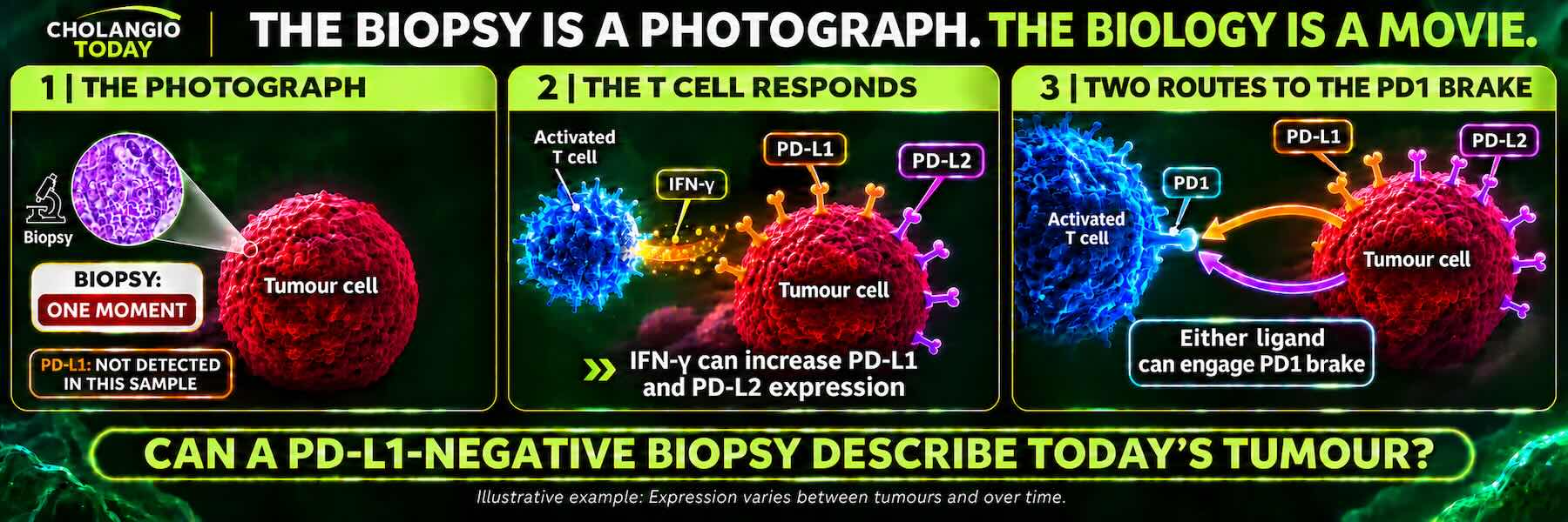
When an Immune Attack Applies Its Own Brake
Return for a moment to Cell City.
Imagine a T cell recognising a cancer cell as a threat and beginning an attack.
As part of that response, activated T cells release a chemical messenger called interferon-gamma, or IFN-γ. Other immune cells, including natural killer cells, can release it too.
Think of interferon-gamma as an alert travelling through the Cell Cities that make up the tumour.
The message tells nearby cells that immune activity is taking place. Cells equipped to receive that message respond by changing their behaviour, including which proteins they produce.
One of those changes can be an increase in PD-L1.
This is not speculation. Interferon-gamma does induce PD-L1 expression in responsive cells. The mechanism has been demonstrated.
And here is where the story becomes particularly important.
PD-L1 can bind to PD1 on T cells and apply an inhibitory braking signal, reducing their activity.
Follow what has happened.
The immune system recognises a threat and begins an attack. Activated immune cells release interferon-gamma. Responsive cells increase PD-L1 expression. That PD-L1 can then engage PD1 and restrain the immune cells carrying out the attack.
The attack has helped activate a brake against itself.
Scientists call this adaptive immune resistance.
But the name is less important than understanding what happens.
The immune system can trigger a response in the tumour environment that makes its own attack harder to sustain.
The immune attack can activate the very brake that restrains it.
For me, this changed the way I understood PD-L1.
It was not simply a protein that a tumour either possessed or did not possess. Its expression could be influenced by the immune activity taking place around it.
That raised an immediate question.
If immune activity can increase PD-L1, what happens when that activity changes?
What the Researchers Had Already Found
I wanted to know how much of this biology had actually been observed.
The research was there.
Taube and colleagues had investigated the relationship between PD-L1 expression and inflammatory immune activity in melanoma. Their work showed how PD-L1 expression could appear alongside an active immune response, supporting the mechanism of adaptive immune resistance.
Spranger and colleagues examined how CD8 T cells could influence the tumour environment, including the development of mechanisms that suppress immune activity.
Vilain and colleagues examined tumour tissue collected before and during PD1 treatment. Their findings documented changes in immune-cell infiltration and PD-L1 expression as treatment and immune activity unfolded.
These studies examined different aspects of the same larger biological picture.
They showed that the tumour environment responds to immune activity and that PD-L1 expression can change in response to it.
This was a significant observation.
A PD-L1 result is often presented as though it describes a settled characteristic of the cancer.
But the evidence showed that PD-L1 expression can be influenced by an active biological process.
The test may accurately measure what is present at the time.
It does not establish that the same conditions existed previously or will remain unchanged.
That distinction led me to a question I had not seen adequately answered by the original response figures.
What Happens When the Immune Attack Weakens?
We had established that an active immune response can increase PD-L1 expression.
Now consider what happens when the immune response itself begins to weaken.
T cells operating within a tumour can face prolonged stimulation, repeated exposure to cancer cells and conditions that interfere with their ability to function.
Over time, some undergo biological changes that reduce their effectiveness.
This is commonly called T-cell exhaustion.
The word can be misleading. These cells are not simply tired in the everyday sense. Their internal programmes change, impairing important functions and making them less responsive. Some become less capable of producing the chemical signals and carrying out the attacks needed to control cancer.
This is established biology.
Now return to interferon-gamma.
If the activity of the immune cells producing interferon-gamma declines, the amount of that signal reaching surrounding cells may also decrease.
And where PD-L1 expression has been increased by interferon-gamma, reduced signalling can reduce that particular source of stimulation.
This does not mean PD-L1 must disappear.
Other mechanisms can maintain or increase its expression. Different cells respond differently, and the tumour environment contains many interacting controls.
But the biological relationship is clear.
Immune activity can influence PD-L1 expression. Changes in that activity can change the signals that regulate it.
The next question followed naturally.
Could a tumour that once experienced a stronger immune attack later show low PD-L1 expression because the immune environment had changed?
The question did not arise from imagination. It arose from putting established observations together.
What Might Have Happened Before the Biopsy?
Imagine following a tumour through time rather than examining it at a single moment.
An immune response develops. T cells recognise cancer cells and begin attacking them.
Those activated T cells release interferon-gamma.
Responsive cells in the tumour environment increase PD-L1 expression, introducing an inhibitory braking signal through PD1.
As the interaction continues, persistent stimulation and suppressive conditions can weaken parts of the T-cell response.
If interferon-gamma signalling subsequently declines, some of the PD-L1 expression maintained by that signalling may also decrease.
Then a biopsy is taken.
The laboratory examines the tissue and classifies it as PD-L1 negative.
The result may be entirely accurate.
But what has it told us about the immune response that occurred before the biopsy?
Very little.
It has not shown us whether the tumour previously experienced a stronger immune attack. It has not reconstructed the earlier chemical signals between the cells. It has not established whether PD-L1 expression was previously higher.
And it cannot tell us whether those conditions might change again.
This is the distinction that matters.
The individual biological events are supported by research. Their possible sequence raises a question about what a single negative result might leave unseen.
What we do not know is whether that sequence occurred in the patients who responded in CheckMate 067.
The published response percentages do not reconstruct their earlier immune histories.
But missing evidence about their earlier biology does not erase the responses that were actually observed.
That is precisely why the question deserves to remain open.
The Evidence Was Pointing Beyond One Explanation
The earlier chapter had established the boundaries of a negative test: sampling, scoring and the fact that PD-L2 is not measured by a standard PD-L1 assay.
Now there was a different dimension. Immune activity can induce PD-L1 expression, meaning what is detectable may change as biological conditions change.
These are distinct limitations, not competing explanations. Together, they show why a negative classification can be accurate without describing the tumour's entire checkpoint environment or its history.
None establishes which mechanism explains the CheckMate 067 responders. But the changing biology makes it harder to justify treating one measurement as a permanent biological verdict.
What Would I Want the Researchers to Investigate?
By this stage, I had a clearer picture of what remained unanswered.
If patients classified as PD-L1 negative responded to nivolumab, what was happening in their tumours that the baseline PD-L1 classification had not captured?
Had some experienced stronger immune activity before the biopsy?
Did PD-L1 expression differ between tumour regions?
Were surrounding immune cells expressing PD-L1?
Was PD-L2 contributing to inhibitory signalling through PD1?
Did the immune environment change during treatment?
These are not questions that require us to disregard the clinical trials.
They arise because of what the trials demonstrated.
The next step would be to examine the available evidence more deeply, including tissue collected at different times, the distribution of immune cells, the expression of PD-L1 and PD-L2, and other measures of immune activity.
Some investigations may confirm an explanation. Others may rule one out or reveal something we have not yet considered.
That is how understanding advances.
I am not seeking an explanation that supports a preferred conclusion.
I am seeking the explanation that survives examination of the evidence.
And until that explanation is established, the unanswered question should remain visible.
Especially when the answer could affect a patient's treatment opportunities.
Why This Matters to the Patient
Imagine being told that your cancer is PD-L1 negative.
That may accurately describe the result of a particular test. But if the explanation goes further, treating it as proof that the PD1 pathway has no relevance or that PD1 treatment could not possibly work, it exceeds what the evidence establishes.
The changing biology adds a reason for caution. PD-L1 expression can respond to immune activity; a result measured at one moment does not establish what happened earlier or what may happen later.
That does not guarantee that PD1 treatment will benefit a patient with cholangiocarcinoma.
The next step is to ask what the result means for the specific treatment being considered, what other biomarkers and clinical evidence are available, and whether the decision has been reviewed with appropriate expertise.
A patient should not have to become a scientist to ask those questions, or accept a conclusion that goes beyond the evidence.
My responsibility is to make that distinction visible early enough for it to matter.
The Question Now Reaches Cholangiocarcinoma
The biology itself could change.
An immune attack can induce PD-L1 expression. That expression can contribute to restraining the attack. Changes in immune activity can alter the conditions regulating PD-L1.
A biopsy cannot reconstruct all of that history from one moment in time.
The individual mechanisms are supported by research. Whether that sequence occurred in the patients who responded in CheckMate 067 remains unknown.
The investigation was approaching its most important test.
What did this mean for cholangiocarcinoma, a cancer in which patients may have limited treatment opportunities and little time to explore them?
That was where I needed to take the evidence next.
A negative PD-L1 snapshot cannot tell us the immune system's history, or determine its future response.
07 | Cholangiocarcinoma
What Does It Really Mean When a Tumour Is Called Immune-Cold?
My investigation was never about melanoma alone.
I had survived Stage IV cholangiocarcinoma. I knew what it meant to confront advanced disease when conventional treatment opportunities had ended and time seemed to be running out.
Understanding was not simply important. It was critical.
Now the investigation returned to the cancer that had changed my life. Cholangiocarcinoma is frequently described as an immune-cold cancer. What did that actually mean? Was it an explanation of why the immune system could not attack, or a description of what researchers observed at a particular time? And were tumours born cold, or could they become cold, and then colder, as their defences developed?
What Does Immune-Cold Actually Mean?
Return to Cell City. A tumour is not simply a mass of cancer cells. It contains blood vessels, supporting tissue, immune cells and other healthy cells interacting with the cancer.
For an effective immune attack, T cells must recognise something abnormal about the cancer, reach their targets and remain capable of attacking them.
In many cholangiocarcinomas, those steps are difficult. Researchers use immune-cold to describe limited effective anti-cancer immune activity. But the label does not identify which obstacle is responsible.
A tumour with few T cells because the immune system has not effectively recognised it faces a different problem from one where T cells recognise the cancer but cannot enter. Both differ from a tumour in which T cells arrive but are restrained.
One label can conceal several different biological problems. A patient cannot act on a label alone.
The Scar-Like Barrier Around the Cancer
One characteristic of cholangiocarcinoma is the dense supporting tissue that can develop around cancer cells. Researchers call it stroma.
Think of what happens when you cut your skin. Your body recognises the injury and begins repairing the damaged area. Specialised cells produce collagen and other materials that rebuild and strengthen tissue. As healing progresses, scar tissue forms.
Cancer cells and surrounding inflammatory signals can also stimulate specialised cells called fibroblasts to produce collagen and supporting materials. Unlike an ordinary wound, where repair eventually settles, a tumour can keep driving this response. The result can be a dense accumulation of scar-like tissue around and between cancer cells.
Think of it as a wound-repair system that has become part of the tumour's environment. This does not mean the cancer began as a physical wound. Cancer can activate wound-repair-like processes without an ordinary injury.
Stroma influences blood vessels, immune-cell movement, chemical signals and treatment response. In some circumstances, this dense environment makes it harder for attacking T cells to reach cancer cells.
Imagine police officers in Cell City trying to reach a dangerous building, only to find the streets blocked. They may recognise the threat and be ready to respond, but cannot reach the target.
That is an access problem, not necessarily a recognition problem. Releasing an immune brake does not automatically remove the physical and chemical barriers preventing the immune cells from reaching cancer.
Are Tumours Born Cold, or Do They Become Colder?
This question changed how I viewed the label.
Cholangiocarcinoma does not necessarily begin with every immune barrier already established. As cancer develops, it interacts with surrounding healthy cells, recruits supporting cells, influences blood vessels and changes chemical signals.
Fibroblasts can produce more scar-like material. Suppressive immune cells can accumulate. T cells can become excluded from cancer-containing regions or lose important functions. Cancer can develop additional ways to escape immune attack.
Some tumours have weak immune recognition early in their development. Others encounter immune pressure and acquire further ways to resist it. There is no single rule that every cholangiocarcinoma becomes progressively colder. Different barriers can change in different directions.
But the evidence establishes that the immune environment develops and changes, and that additional barriers can emerge.
If the tumour is building defences, why are we not investigating treatment opportunities before those defences become more established?
This makes timing part of the biological investigation. A patient may be fit for a treatment today but too unwell to receive it later. A tumour may be more accessible to immune cells under one set of conditions than another. The opportunity may depend on both the intervention and when it is delivered.
We do not yet know that earlier PD1 treatment prevents immune suppression in cholangiocarcinoma. But the changing biology gives us a reason to investigate timing, rather than waiting until the patient has exhausted every conventional option before beginning the discussion.
Three Different Problems: Recognition, Access and Activity
The evidence became easier to understand when I separated three questions.
Can the immune system recognise the cancer? Cancer cells can contain abnormal proteins that distinguish them from healthy cells. Some of those abnormalities can be presented to T cells, but recognition is not equally strong in every tumour.
Can attacking cells reach the cancer? Dense supporting tissue, abnormal blood vessels and chemical signals can interfere with entry and movement.
Once they arrive, can they remain active? T cells may encounter PD1 checkpoint signalling, suppressive immune cells, low oxygen, limited nourishment or prolonged stimulation that impairs important functions.
These are different biological obstacles. A PD1 inhibitor addresses an inhibitory signal through the PD1 receptor. It does not automatically create recognition, open every route into the tumour or remove every suppressive condition.
Understanding which obstacle is present matters more than repeating the description immune-cold.
Can We Disrupt an Immune-Cold Tumour?
Another question emerged as I examined those barriers.
If a tumour develops an environment that protects it from immune attack, can we intervene to disrupt that environment?
Radiation, TACE and emerging technologies such as histotripsy approach the tumour differently. Radiation damages cancer cells using high-energy radiation. TACE delivers treatment through arteries supplying the tumour and reduces its blood supply. Histotripsy uses focused sound waves to mechanically destroy targeted tissue.
Beyond their direct effects, these interventions can alter the surrounding biological environment. Damaged cancer cells can release material the immune system may recognise. Inflammatory signals can change. Immune cells may be recruited or activated.
Think of Cell City. An intervention disrupts part of the environment in which the cancer has established its defences.
Could that disruption help the immune system recognise and attack cancer cells more effectively? And if immune activity increases, could PD1 treatment prevent PD-L1 and PD-L2 from applying their inhibitory brakes?
Researchers are investigating combinations of local tumour treatments and checkpoint medicines. Early cholangiocarcinoma studies, including radiation combined with PD1 treatment, have reported responses. Those responses do not yet prove that the intervention converted an immune-cold tumour into one responsive to PD1. Disruption can also create inflammatory or suppressive effects that work against immunity.
The question is which intervention produces useful immune changes, for which patients, and when.
Could the opportunity for PD1 treatment depend not only on which medicine we use, but on what we do to the tumour before or alongside it?
That is an inference worth making visible to patients and their doctors. Evidence establishes mechanisms; observation connects them; the next task is to test whether the connection improves outcomes. The question itself can change what a patient and doctor investigate while treatment remains possible.
What Can DNA Repair Tell Us About Immune Recognition?
Our cells contain DNA, the instructions used to build and maintain the body. When DNA is copied, mistakes can occur. Cells have repair systems that correct many of them.
One is mismatch repair, or MMR. Think of the DNA instructions in Cell City's Library. The mismatch repair system acts like a proofreader, checking for certain copying errors. When it is deficient, called dMMR, errors can remain and accumulate.
Microsatellites are short sequences of DNA letters repeated several times. Imagine a line copied as:
STOP STOP STOP STOP
A copying error might change the number of repetitions:
STOP STOP STOP
Or add another:
STOP STOP STOP STOP STOP
The words are recognisable, but the number of repetitions has changed. This illustrates what microsatellite instability testing detects: changes in repeated DNA sequences. Such changes do not necessarily alter a protein instruction themselves. They are evidence that copying and repair have become unreliable.
Some DNA errors, including errors arising with deficient mismatch repair, can produce abnormal proteins that the immune system may recognise. An MSI-high result indicates substantial instability at the DNA markers examined. dMMR and MSI-high can be important when assessing certain immunotherapy opportunities.
But MMR, MSI and PD-L1 are not the same measurement. A cancer without dMMR or MSI-high may still contain abnormal proteins that the immune system recognises. And immune recognition does not guarantee high PD-L1 staining in a particular biopsy.
Different biological questions require different measurements.
What Does Immune-Cold Not Tell Us?
By now, immune-cold had become less of an answer and more of a starting point.
It could not, by itself, tell me whether the main obstacle was recognition, access or activity, or whether those conditions might change.
Calling a tumour immune-cold does not identify every barrier or establish that no intervention could alter one.
That was the distinction I wanted to bring into the patient's understanding.
What Should Patients and Doctors Ask Earlier?
When a tumour is described as immune-cold, the first question is what that label actually tells us about this particular cancer.
Has the medical team identified evidence of immune recognition? Are T cells present in or around the tumour? Is there evidence that they are excluded or suppressed? Have relevant biomarkers, including MMR and MSI where appropriate, been assessed?
Could a local intervention change the conditions preventing an immune attack? Would that make an immune treatment or a clinical trial worth considering? And, crucially, when must those questions be answered before the opportunity disappears?
Not every patient needs every test or intervention. But patients and their doctors should be able to distinguish between what has been measured, what is inferred and what remains unknown.
The earlier right action is to identify the actual barrier, investigate whether it can be addressed and do so while the patient still has options.
The Question Becomes Personal
I was not trying to establish that every patient with cholangiocarcinoma should receive PD1 treatment. I was investigating whether patients were being given a complete account of the evidence before a treatment opportunity was dismissed.
Patients may already have received chemotherapy and immunotherapy. Their cancer may have progressed. When they ask about another immune checkpoint medicine, they may hear that they have already tried immunotherapy.
But had they tried the same biological intervention?
That was where the investigation needed to go next.
When a tumour is building defences, the time to investigate how to overcome them is before the patient's opportunities disappear.
08 | The Treatment Difference
Has Durva Really Closed the Door on Keytruda and Nivo?
Progression after Durva establishes that one treatment strategy has not maintained control of the cancer. It does not, by itself, establish that PD1 treatment has been exhausted.
Imagine a patient with advanced cholangiocarcinoma who has received gemcitabine, cisplatin and durvalumab, commonly known as Durva.
For a time, the treatment may have controlled the cancer. Then a scan reveals progression.
The patient asks whether pembrolizumab, known as Keytruda, or nivolumab, known as Nivo, might still be worth investigating.
The answer may be that the patient has already tried immunotherapy.
I wanted to understand what that answer actually established.
Durva, Keytruda and Nivo belong to the same broad family of checkpoint inhibitors. They interfere with mechanisms that can restrain the immune system's ability to attack cancer. But they are not the same medicine, and they do not block the same part of the checkpoint system.
That difference brought me to a question.
If Durva has failed to maintain control of the cancer, what evidence establishes that Keytruda or Nivo cannot work?
One Receiver. Two Ways to Apply the Brake.
Earlier in this investigation, we entered Cell City to understand how the immune system recognises cancer and how cancer can interfere with that response.
Imagine the T cells as Cell City's police. They are trained to recognise threats, but they also carry safety equipment designed to prevent unnecessary damage to healthy tissue.
One of those safety controls is PD1, a receptor on the T cell.
Think of PD1 as a receiver capable of accepting an instruction to apply the brake.
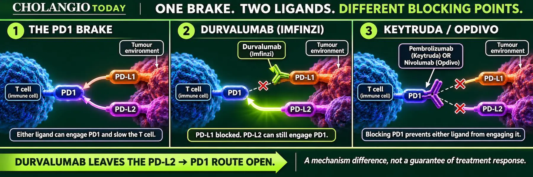
Two proteins, PD-L1 and PD-L2, can connect to that receiver and deliver inhibitory signals.
Cancer can use this normal protective system to weaken an immune attack.
Durva binds to PD-L1. By doing so, it prevents PD-L1 from engaging PD1 and delivering its inhibitory signal. But it does not block PD-L2 from engaging PD1.
Keytruda and Nivo work differently. They bind to the PD1 receptor itself, preventing both PD-L1 and PD-L2 from engaging that receptor.
In Cell City, Durva intercepts one of the messages telling the police to stand down. Keytruda and Nivo block the receiver through which either message would be delivered.
The distinction is established biology.
It does not tell us that one treatment will necessarily succeed where another has failed. It tells us that the treatments are biologically different.
And that difference matters when we examine what a patient's previous treatment has actually established.
What Has Actually Failed?
When a patient's cancer progresses after gemcitabine, cisplatin and Durva, we know that the treatment strategy has not maintained control of the disease.
We do not know, from that observation alone, which biological factors account for the progression.
The cancer may have changed. The immune cells may be unable to recognise it effectively. The tumour environment may be preventing those cells from reaching or attacking the cancer. Other inhibitory pathways may be involved.
The treatment also contains chemotherapy, which has its own effects on cancer cells and the surrounding immune environment.
Progression therefore establishes an outcome of the treatment given. It does not establish the outcome of every treatment that shares one of its characteristics.
Durva blocks PD-L1. Keytruda and Nivo block PD1.
If the patient has received Durva but not Keytruda or Nivo, those medicines have not been tested in that individual simply because all three belong to the broader family of checkpoint inhibitors.
I wanted to see whether the clinical trials had answered the question that mattered to this patient.
Durva on Its Own
Before examining the larger combination trials, I discovered that Durva had also been investigated on its own.
An early-phase study involving patients in Asia examined Durva monotherapy in people with advanced biliary tract cancer whose disease had progressed during or after chemotherapy treatment.
Among the 42 patients with biliary tract cancer who received Durva alone, the reported objective response rate was 4.8%.
That meant responses had been documented without concurrent chemotherapy.
The same study also investigated Durva combined with tremelimumab, a medicine that blocks CTLA-4. In that separate group of 65 patients with biliary tract cancer, the objective response rate was 10.8%.
These were different treatment groups, not a randomised comparison establishing that one approach was superior.
But each result was an observation worth retaining.
Durva alone had produced responses. Durva combined with CTLA-4 blockade had also produced responses.
The next question was what happened when checkpoint treatment was combined with chemotherapy.
TOPAZ-1 and KEYNOTE-966: What Did They Actually Test?
Two major trials helped establish checkpoint inhibitors alongside chemotherapy as first-line treatment options for advanced biliary tract cancer.
TOPAZ-1 investigated Durva combined with gemcitabine and cisplatin.
KEYNOTE-966 investigated Keytruda combined with gemcitabine and cisplatin.
Both trials compared their respective combinations against chemotherapy-based control treatment.
Both demonstrated improvements in overall survival with the addition of checkpoint treatment.
In TOPAZ-1, median overall survival was 12.8 months with Durva plus chemotherapy, compared with 11.5 months in the control group.
In KEYNOTE-966, median overall survival was 12.7 months with Keytruda plus chemotherapy, compared with 10.9 months in the control group.
Those figures came from separate trials. They were not a head-to-head comparison between Durva and Keytruda.
The earlier monotherapy studies had already documented clinical responses to checkpoint blockade. The later combination trials demonstrated survival improvements when checkpoint inhibitors were added to chemotherapy.
Both approaches produced evidence, but they answered different questions.
And the question that interested me was not which trial reported the slightly higher median survival.
It was what the trials had actually tested.
TOPAZ-1 tested the addition of Durva to a chemotherapy-based treatment strategy.
KEYNOTE-966 tested the addition of Keytruda to a chemotherapy-based treatment strategy.
Neither trial directly tested whether Keytruda or Nivo could produce a response in patients whose cancer had progressed after Durva.
Neither trial isolated the effect of PD1 monotherapy in that post-Durva setting.
The trials answered important questions about the treatment combinations they investigated.
They did not answer every question about the individual medicines or their possible use in a different sequence.
What Was Chemotherapy Doing to the Immune System?
This led me to another question.
Checkpoint inhibitors depend upon the patient's immune system.
Chemotherapy acts through different mechanisms, including interference with the processes that dividing cells need to survive and reproduce.
Cancer cells are among its targets, but healthy dividing cells can also be affected. These include cells involved in immune function.
Chemotherapy can damage healthy cells and reduce some immune-cell populations, including T cells. That is a biological effect worth examining when the treatment being combined with chemotherapy depends upon those same immune cells.
Chemotherapy can damage healthy cells too, including immune cells such as T cells, the very cells checkpoint treatment depends upon.
This left me with another question. Chemotherapy is intended to damage cancer cells, but could it also hinder the T cells that checkpoint treatment is attempting to release from inhibition?
Could a treatment intended to weaken the cancer also weaken the immune response needed to fight it?
Yet the relationship is not simply one of suppression.
Certain chemotherapy treatments can also make cancer cells more visible to the immune system, change the tumour environment or stimulate immune responses through the effects of cancer-cell damage.
That means chemotherapy may influence checkpoint treatment in more than one direction.
I could not assume that chemotherapy necessarily weakened the overall effect of Keytruda or Durva.
Nor could I assume that combining chemotherapy with a checkpoint inhibitor revealed everything that the checkpoint inhibitor might do when given without concurrent chemotherapy.
The combination trials had measured the effects of the combinations.
They had not separated every contribution of the medicines or every consequence of their interaction with the immune system.
Think again of Cell City's police.
A checkpoint inhibitor may remove an instruction telling them to stand down. Chemotherapy may simultaneously change the number, condition and surroundings of those officers, while also changing the threat they are attempting to recognise.
What happens when these interventions are combined is a legitimate experimental question.
What happens when they are separated is another.
They are not automatically the same question.
What If the Unanswered Question Matters More Than We Recognised?
The more I examined these trials, the more I began to question not only what the results showed, but what the experiments had left unanswered.
The results were what they were. I had no reason to dispute them.
But the results reflected the questions the scientists had chosen to investigate, the treatments they had chosen to combine and the conditions under which they had chosen to measure them.
Other questions remained unanswered.
The earlier PD1 studies had documented responses without concurrent chemotherapy, including in patients classified as PD-L1 negative. The later combination trials investigated checkpoint treatment alongside gemcitabine and cisplatin, medicines that also affect the immune cells upon which checkpoint treatment depends.
Both approaches produced evidence. But they did not answer the same biological question.
As my understanding grew, so did my doubt.
Not doubt about the reported results, but about the completeness of the answers being carried forward.
Science advances through what we discover. Yet every discovery also sits alongside what we have not discovered, what we have not tested and what we may not yet recognise.
Scientists decide which questions to pursue. Those choices shape the evidence that becomes available, but they do not determine the limits of what is biologically possible.
What if the questions left unanswered were as important as those answered?
And what if the conclusions being carried forward had become broader than the evidence itself?
For the patient who has exhausted conventional treatment options, that distinction matters enormously.
A trial may establish what a particular treatment achieved. It cannot establish the failure of a different treatment it never tested.
The more I understood, the clearer that distinction became.
Science contains both the known and the unknown. An answer is only complete when it makes clear what it establishes and what remains unanswered.
What About CTLA-4 and PD1 Together?
There was another treatment difference I needed to understand.
Nivo blocks PD1.
Ipilimumab, known as Ipi, blocks CTLA-4, a different immune checkpoint that normally restrains T-cell activation.
Combining Nivo and Ipi therefore interferes with two distinct mechanisms that can restrain an immune response.
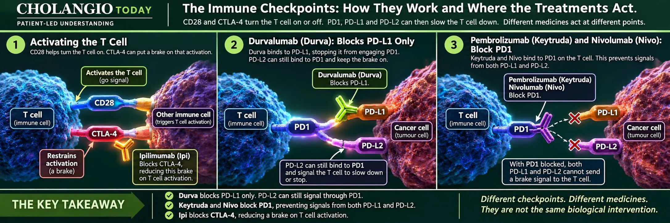
The combination is not simply another name for Durva or Keytruda.
In CheckMate 067, a trial involving advanced melanoma, responses were documented in patients classified as PD-L1 negative who received Nivo alone and in those who received Nivo together with Ipi.
The combination produced a higher response rate than Nivo alone in that PD-L1-negative melanoma subgroup, although it also carried substantially greater risks of serious immune-related adverse effects.
That observation cannot be transferred into a predicted response rate for cholangiocarcinoma.
But it demonstrates something relevant to the investigation: a negative PD-L1 classification did not prevent responses to PD1 blockade, including when PD1 and CTLA-4 were blocked together.
Studies in advanced biliary tract cancer have also documented responses with combined Nivo and Ipi.
These are different treatments, investigated under different conditions.
Their outcomes deserve to be examined individually rather than dismissed under the single description of immunotherapy.
The Evidence Already Exists
The melanoma trials had already established that patients classified as PD-L1 negative could respond to PD1 blockade. Their results could not predict outcomes in cholangiocarcinoma.
More importantly for this investigation, evidence also existed in advanced biliary tract cancer.
KEYNOTE-158 investigated Keytruda without concurrent chemotherapy in previously treated patients with advanced biliary tract cancer.
Six of the 104 patients in the reported cohort experienced an objective response.
Among the 34 patients classified as PD-L1 negative in the reported subgroup, one experienced a response.
That was a small number, but it was a documented clinical response.
The published subgroup result does not, by itself, identify which biliary tract cancer subtype that individual had. It therefore should not be presented as proof of a response in PD-L1-negative cholangiocarcinoma specifically.
Nevertheless, it establishes that a PD-L1-negative classification did not exclude a response to Keytruda in the advanced biliary tract cancer population studied.
The evidence was not a theory.
The patients had been treated. Their responses had been measured and reported.
The unanswered questions concerned how to interpret those observations, what they meant for different patient groups and treatment sequences, and which opportunities remained to be investigated.
They did not erase the observations themselves.
The Decision Window Opens Before Durva Fails
For a patient with advanced cholangiocarcinoma, time matters.
A person may be well enough to consider further treatment when progression is first recognised, but that opportunity can narrow as the disease advances.
That makes the timing of understanding important.
Before first-line treatment fails, patients can begin asking what their tumour testing has established, which biomarkers have been examined, whether comprehensive genomic testing has been completed and which treatment or clinical-trial opportunities might become relevant.
They can also ask about the differences between Durva, Keytruda, Nivo and combined checkpoint strategies.
The purpose is to prevent a potentially relevant question from being discovered only after the patient has lost the opportunity to investigate it.
In Australia, access to a treatment outside its approved or funded indication may require additional clinical assessment and a lawful access pathway. Those practical requirements matter, and they take time.
Earlier understanding gives the patient and their treating team more time to establish what is known, what remains uncertain and what action is possible.
When Conventional Options Have Run Out
Now return to the patient whose cancer has progressed after Durva.
The patient may have exhausted established treatment options. Their PD-L1 result may be negative. They may be told that further immunotherapy is not an option because immunotherapy has already failed.
But what has the evidence actually established?
A negative PD-L1 classification did not prevent responses to PD1 treatment in the studies we have examined.
Durva and PD1 inhibitors act at different points in the checkpoint pathway. Their clinical results have also been measured in different trials, under different treatment conditions.
And progression after Durva does not, by itself, establish that Keytruda, Nivo or a different checkpoint strategy cannot work.
That does not mean the precise post-Durva treatment sequence has been demonstrated to be effective in cholangiocarcinoma.
It means the outcome of that untested sequence cannot be declared from the failure of a different treatment.
For the patient, the consequence is clear.
A negative PD-L1 result or progression after Durva cannot be interpreted as proof that PD1 treatment cannot work.
The patient has every reason to ask that the relevant evidence be examined, the treatment differences understood, and the remaining lawful options considered before an opportunity is closed.
When the Patient Is Told the Door Has Closed
There is a question I would want every patient to feel able to ask when a treatment opportunity is dismissed.
What evidence establishes that exclusion?
Not whether the treatment is routine.
Not whether it is funded.
Not whether a guideline has already adopted it.
Those are separate questions, and each may matter when determining what can actually be done.
But they do not change the findings of a clinical trial or turn an unanswered biological question into a negative result.
The clinician and the patient must work from the same evidence.
The clinician brings medical knowledge, experience and responsibility for safe treatment. The patient brings the full consequence of the decision and the right to understand what is being decided.
Neither should have to pretend that the evidence establishes more than it does.
The patient needs to know which treatments have demonstrated responses, which circumstances have been tested, which have not, and what remains possible to investigate.
That is the difference between explaining the limits of evidence and allowing those limits to become an unsupported exclusion.
The Question That Remains
I began this chapter with a patient whose cancer had progressed after Durva.
The patient wanted to know whether Keytruda or Nivo might still be worth investigating.
The answer could not be found simply by saying that the patient had already received immunotherapy.
We had to examine the biological differences between the medicines, the experimental questions answered by the trials and the clinical responses that had already been observed.
That examination brought us back to the finding at the heart of this investigation.
PD1 treatment had produced documented responses in patients classified as PD-L1 negative, including a response in the PD-L1-negative advanced biliary tract cancer subgroup of KEYNOTE-158.
The evidence remained intact.
The unanswered questions did not diminish it. They showed us where further investigation was needed and where conclusions must not extend beyond what had actually been tested.
The next chapter returns to the clinical evidence in biliary tract cancer and asks what those observations mean for the patient who needs to make a decision while there is still time.
A treatment family is not a single treatment. Durva progression does not, by itself, close the door on PD1.
09 | The Evidence and the Opportunity
What Have Clinical Trials Already Revealed About PD1 Treatment in Biliary Tract Cancer?
Chapter 08 had established that Durva, Keytruda and Nivo are not identical biological interventions. It had also revealed an unanswered question about what happens when one checkpoint treatment is followed by another.
But there was something more important than the difference between the medicines.
What had already happened when patients with biliary tract cancer received PD1 treatment?
I wanted to follow the clinical evidence into the disease itself.
Not to predict what would happen to the next patient, but to establish what researchers had already observed in patients who had received these treatments.
The evidence was there.
KEYNOTE-158: The Observation Reaches Biliary Tract Cancer
KEYNOTE-158 investigated pembrolizumab, known as Keytruda, in patients with previously treated advanced biliary tract cancer.
Among 104 patients, six experienced an objective response.
That represented 5.8% of the study population.
The overall response rate was low. Most patients did not achieve an objective response.
But within those results was a finding of particular importance to this investigation.
Thirty-four patients had been classified as PD-L1 negative.
One of those 34 patients responded to Keytruda.
That represented 2.9% of the PD-L1-negative subgroup.
One patient.
A small number, certainly. But this was not an imagined biological possibility. It was a clinical response observed, measured and reported in a patient with advanced biliary tract cancer.
The finding had now moved beyond melanoma.
The published subgroup results did not identify the particular biliary tract cancer subtype of that responder. That detail remained unanswered.
The observation itself did not.
A patient classified as PD-L1 negative had responded to a medicine blocking PD1.
I wanted to understand something else about those responses.
How long did they last?
What the Response Rate Did Not Reveal
An overall response rate tells us how many patients achieved measurable tumour shrinkage meeting the study's response criteria.
It does not, by itself, describe the experience of those patients over time.
In KEYNOTE-158, some responses were durable. The published study reported response durations extending from at least 6.2 months to more than two years.
That added another dimension to the finding.
A response lasting a few months and one continuing for years both contribute to the response rate, but they can have very different consequences for the patient.
The six responses were not evidence that most patients benefited through measurable tumour shrinkage. They were evidence that some patients did respond, and that some responses persisted.
For a patient searching for another opportunity, understanding both the frequency and the duration of responses matters.
The study also examined microsatellite instability, or MSI.
Among patients whose MSI status was assessed, none were classified as MSI-high, including those who responded.
The responses therefore extended beyond patients with that particular DNA repair-related characteristic.
This was another observation worth retaining.
The investigation was no longer confined to whether PD-L1-negative responses could occur.
It was beginning to reveal clinical responses in biliary tract cancer that could not simply be attributed to MSI-high disease.
I next wanted to know what researchers had observed specifically in cholangiocarcinoma.
Nivolumab: The Evidence Reaches Cholangiocarcinoma
A Phase 2 study led by Kim and colleagues investigated nivolumab, known as Nivo, in patients with previously treated advanced biliary tract cancer.
Unlike a single overall response figure, the investigators also reported outcomes according to where the cancer began.
Among 28 patients with intrahepatic cholangiocarcinoma, which begins in the bile ducts inside the liver, six experienced an objective response.
Among five patients with extrahepatic cholangiocarcinoma, which begins in the bile ducts outside the liver, two experienced an objective response.
These were patients with cholangiocarcinoma.
They had received Nivo.
And clinical responses had been documented.
Across the 46 patients evaluable for response, investigators reported ten objective responses, representing approximately 22%.
An independent central review reported five responses, representing approximately 11%. One response in each assessment was unconfirmed.
The two assessments produced different results. Both belong in an accurate account of the trial.
What mattered to my investigation was the clinical observation they shared.
Nivolumab had produced measurable responses in patients with cholangiocarcinoma.
This was not an inference drawn from melanoma.
It was evidence from patients with the disease I was investigating.
And it led to another question.
If blocking PD1 could produce responses in cholangiocarcinoma, what had researchers observed when they blocked PD1 and CTLA-4 together?
Two Checkpoints: Another Clinical Observation
The discoveries of Allison and Honjo had revealed two different controls on immune activity.
CTLA-4 helps regulate T-cell activation. PD1 can restrain activated T cells as they respond.
Nivo blocks PD1.
Ipilimumab, known as Ipi, blocks CTLA-4.
Together, they interfere with two distinct immune restraints.
Researchers had investigated this combination in advanced biliary tract cancer.
The CA209-538 study documented responses among patients receiving Nivo and Ipi.
The Australian MoST-CIRCUIT trial subsequently investigated the same combination in patients with intrahepatic cholangiocarcinoma and gallbladder cancer.
These studies added another clinical observation to the investigation.
Responses had been documented with combined checkpoint blockade in biliary tract cancers.
The treatment was different from Nivo alone, Keytruda alone or Durva combined with chemotherapy.
And it had its own clinical evidence.
I was beginning to see the importance of examining each treatment according to what had actually been administered and observed.
The medicines might belong to the same broad family, but their biological targets and treatment combinations were not identical.
Neither were the questions their trials had answered.
What the Evidence Had Established
I had followed the investigation from melanoma into biliary tract cancer and then into cholangiocarcinoma itself.
The findings were no longer confined to one observation.
KEYNOTE-158 had documented responses to Keytruda in advanced biliary tract cancer, including a patient classified as PD-L1 negative.
The study led by Kim and colleagues had documented responses to Nivo in patients with intrahepatic and extrahepatic cholangiocarcinoma.
CA209-538 and MoST-CIRCUIT added clinical evidence involving combined PD1 and CTLA-4 blockade in biliary tract cancers.
Each study contributed its own findings.
Taken together, they established that clinical responses to PD1-based treatment had occurred in these cancer populations.
That was what the evidence said.
I did not need to add an explanation the researchers had not established. Nor did I need to fill gaps in the published findings.
Those gaps remained questions.
What distinguished the patients who responded?
Which biological conditions had enabled their immune systems to attack the cancer?
Could a better understanding of those conditions help identify further treatment opportunities?
And what happened when a patient had already received Durva before another PD1 treatment was considered?
The studies had not answered every question.
But they had provided something essential for the next stage of the investigation.
Documented clinical responses.
When the Evidence Reaches the Patient
I now wanted to move from the research findings to the place where their meaning could become most consequential.
The consultation room.
Imagine a patient with advanced cholangiocarcinoma whose conventional treatment opportunities are running out.
The patient and their oncologist are considering what remains.
The evidence we have examined does not decide that patient's treatment. It provides clinical findings that deserve to be understood as part of the decision.
There is a difference between knowing that a treatment has produced responses and understanding whether it is a reasonable opportunity for a particular patient.
That is where clinical expertise, the patient's circumstances and the complete available evidence must come together.
The patient should not have to undertake this investigation from the beginning simply to discover findings that researchers have already published.
Those findings should be available, understandable and brought forward while there is still time to act.
For me, that was the consequence of following the evidence.
The investigation had begun with a question about patients responding despite a negative PD-L1 classification.
It had now reached documented responses in biliary tract cancer and cholangiocarcinoma.
The next question was no longer simply what the studies had observed.
What should happen when that evidence reaches a patient whose treatment opportunities may be closing or have closed?
That was where the investigation needed to go next.
We do not inflate the few into the many. But neither do we allow the few to disappear because they are few.
10 | When the Evidence Reaches the Patient
What happens when documented clinical evidence meets a patient who needs another treatment opportunity?
The investigation had followed the clinical evidence to a point I could not ignore.
Patients classified as PD-L1 negative had responded to PD1 treatment. Responses had also been documented in biliary tract cancer, including cholangiocarcinoma.
The findings were published. They were available to clinicians, researchers and patients.
But there was another question.
What happens when a patient needs that knowledge?
I have spent years working with patients and families affected by cholangiocarcinoma, often at the most difficult moments of their lives.
Many are newly diagnosed and trying to understand what has happened. Others are responding to progression, investigating another treatment or facing the possibility that conventional options have been exhausted.
Claire and I founded Cholangiocarcinoma Foundation Australia, not for advocacy or awareness, but as a solution. And that solution began with earlier understanding and helping turn that understanding into practical responses. My own experience of surviving Stage IV cholangiocarcinoma shaped that commitment, but it is the years spent working alongside other patients that have shown me how differently a treatment opportunity can look depending on how it is understood and when that understanding is activated. Timing is a crucial component of effective response, no matter how late it may be.
I have seen the value of a question asked early enough to change what happens next.
And I have seen what happens when understanding arrives too late.
That experience is why I wanted to follow this investigation beyond the published clinical trials.
The evidence had reached the scientific literature.
Now it needed to reach the patient.
When the Patient Is Told There Is Nothing More
Imagine a patient with advanced cholangiocarcinoma.
They have received gemcitabine, cisplatin and Durva. Their cancer has progressed.
They may have undergone further chemotherapy, targeted treatment or another available therapy.
Eventually, their oncologist explains that there are no further conventional treatment options to recommend.
For the patient, that conversation can feel like a final judgement.
There may be no more established treatments to offer. But the patient may still be well enough to pursue another reasonable opportunity if one can be identified.
And they have a question.
Has everything that might reasonably help me been considered?
Perhaps they have read this investigation.
They now know that Durva and PD1 inhibitors act on different biological targets at different points in the checkpoint pathway. Their clinical trials have reported different results. They have discovered that PD-L1-negative patients have also responded to PD1 treatment, and that clinical responses have been documented in cholangiocarcinoma.
They bring those findings to their oncologist.
Not to insist that a particular treatment must work.
They want to know what the evidence means for them.
The Evidence Has Arrived. What Happens Next?
A clinical trial records what happened to patients who received a particular treatment under particular conditions.
An oncologist must determine how those findings apply to the person sitting in front of them.
That requires medical judgement.
The patient's current condition, liver function, previous treatment, genomic findings and other relevant test results all contribute to the assessment.
So do the potential benefits and risks of further treatment.
Checkpoint inhibitors can cause serious immune-related complications. Combining PD1 and CTLA-4 blockade can increase those risks.
Another treatment or clinical trial may offer a stronger opportunity.
These considerations are part of making a clinical decision, not reasons to avoid making one.
What matters is that the relevant evidence is examined in the patient's circumstances and the reasoning is explained.
The patient should be able to understand what supports the recommendation, what limits it and what can happen next.
Understanding the Reason for a Decision
Over years of conversations with patients, I have come to appreciate how much can depend on a few words spoken during a consultation.
A patient may hear that a treatment is not recommended.
But what does that mean?
It may mean that the medicine presents an unacceptable medical risk for that patient.
It may mean another available treatment has stronger evidence of benefit.
It may mean that the clinical evidence does not sufficiently support the proposed treatment in their particular circumstances.
Or it may mean that treatment access has not been established.
These are different circumstances, with different consequences.
The distinction matters because a clinical recommendation, a treatment's availability and its biological ability to produce a response are not the same question.
A patient cannot participate meaningfully in a decision without understanding which question has been answered.
If a treatment is not recommended, the patient should know why.
And if the reason reveals another question worth investigating, that question should remain visible.
A Conversation Worth Having
If I were sitting beside a patient who had exhausted conventional treatment, I would want them to have a straightforward conversation with their oncologist.
They should not need to memorise the studies or repeat the biological explanations contained in this investigation.
The evidence can be brought into the consultation.
The patient can then ask:
“Given the published clinical responses to PD1 treatment, including responses in patients classified as PD-L1 negative and in patients with cholangiocarcinoma, is there a reasonable clinical basis to consider Keytruda, Nivo or a PD1 and CTLA-4 combination in my circumstances?”
That question brings the evidence to the decision.
The next question matters just as much.
“If you do not recommend one of these treatments, can you explain the specific clinical reasons, and whether another opportunity would be more appropriate?”
The patient is asking for an individual assessment.
The oncologist brings the clinical expertise to determine what is reasonable and safe. The patient brings their circumstances, their priorities and the consequences of the decision.
Both need to understand the evidence being considered.
And both need to understand what will happen next.
The Patient Should Not Have to Become the Researcher
There is something troubling about the possibility that a patient with advanced cancer might need to discover published clinical evidence before it becomes part of a treatment conversation.
These patients are already managing the physical and psychological demands of their disease, the effects of treatment, uncertainty about their future and the responsibilities of everyday life.
Some have the energy, resources and support to investigate further.
Most do not.
Yet access to understanding should not depend on whether a patient has the capacity to conduct an investigation like this one.
That is why I have undertaken this work.
To identify the observations researchers have already made, understand their significance and carry that knowledge forward to patients who need it, now, today.
It is knowledge that belongs in the clinical conversation, where it can be considered alongside the patient's individual circumstances.
A patient may ultimately receive the treatment they have asked about.
They may receive a different recommendation.
Or the assessment may establish that further treatment is not clinically appropriate.
But the decision should emerge from the available evidence and an examination of the patient's circumstances.
Not from a question that was never asked.
When Understanding Must Become Action
The patient and oncologist have now reached an important point.
The published findings are understood.
The available opportunities have been examined.
A clinical decision can be made.
But for a patient with progressing cholangiocarcinoma, there is one more consideration.
Time.
A treatment that may be reasonable today could become impossible if the patient's condition deteriorates before the decision is made or the medicine can be obtained.
The next stage of the response is therefore more than understanding the evidence.
It is establishing what can be done, how it can be accessed and whether the opportunity can still be pursued.
That is where our investigation must go next.
The evidence must reach the patient, the decision must be understood, and the next action must follow while there is still an opportunity to act.
11 | Before the Opportunity Is Lost
The evidence matters most when understanding it can still change what happens next.
There is a moment in the life of a patient with advanced cancer when time begins to take on a different meaning.
It is no longer simply the number of months since diagnosis, the interval between scans or the date of the next appointment.
Time begins to determine which opportunities remain possible.
A patient may be medically fit enough to consider another treatment today. But cholangiocarcinoma can progress quickly. Deteriorating liver function, infection, complications or declining physical strength may make further treatment increasingly difficult.
For some patients, the next opportunity can be considered while their current treatment is still working.
For others, conventional options have already been exhausted. They may have been told there is nothing further to offer, yet remain well enough to investigate another approach.
Both patients need understanding.
And both need it when they can still do something with it.
What Happens After the Decision?
In Chapter 10, we brought the published evidence into the consultation room.
The patient had asked whether Keytruda, Nivo or a combination involving PD1 and CTLA-4 blockade might offer another reasonable treatment opportunity.
The oncologist's responsibility was to assess that request against the patient's medical circumstances, the available evidence, the risks and any better alternatives.
But even when a treatment is considered clinically reasonable, another problem remains.
How does the patient obtain it?
A medicine cannot help someone who cannot access it.
An appropriate clinical trial may provide a pathway. In other circumstances, access might involve an approved indication, an off-label prescription, a special-access arrangement or a manufacturer-supported programme.
In Australia, a medicine being legally available for prescribing and being funded through the Pharmaceutical Benefits Scheme are different matters.
A treatment may be considered clinically appropriate without being routinely funded for that particular cancer or treatment setting.
And special-access arrangements are not automatic. They require a clinician to assess and support the proposed treatment, with the relevant requirements addressed.
These are practical questions, but they can determine whether a clinical opportunity becomes an actual treatment.
Who will investigate access?
What medical information is needed?
Can the treatment be funded?
Would an experienced specialist offer another perspective?
How long might the process take?
The answers matter because every step consumes time.
For patients whose conventional options have already ended, this work should begin immediately.
For those still responding to established treatment, understanding the next possibilities in advance can provide something valuable: preparation.
An opportunity is not preserved simply because it has been identified. The patient must still be able to pursue it.
The Information We Must Preserve
There is another observation I want to bring forward before concluding this investigation.
It concerns information already available from the patient's tumour.
Durva's biological target is PD-L1.
It is therefore important that PD-L1 expression is tested and recorded in the patient's biopsy results, even though expression levels have not reliably predicted who will benefit from Durva combined with chemotherapy.
PD-L1 testing is not required to prescribe Durva for advanced biliary tract cancer. But that does not make the observation itself unimportant.
My reason for raising this is not to suggest that a PD-L1 result can select every patient who will respond. It is to ask what we might learn if the expression findings, the assay used, the treatment received and the patient's subsequent response were consistently recorded and carried forward across this patient population.
One patient's result is an observation. When comparable observations accumulate across patients, and when further tissue samples are available over time, they may help us discover patterns we cannot yet see.
And there is another question arising from the biology already examined.
PD-L1 is not the only protein capable of applying a brake through PD1.
There is also PD-L2.
Durva blocks PD-L1, but it does not directly block PD-L2. Keytruda and Nivo block PD1, preventing inhibitory signalling through that receptor from both proteins.
A PD-L1 expression result does not reveal PD-L2 expression or its activity. Yet PD-L2 remains part of the pathway, whether or not it appears in the ordinary clinical discussion.
If we are recording one biological brake, what are we learning about the other?
I raise that question not to supply an untested explanation for clinical responses, but because the existence of the second brake gives us reason to remain curious about what a single measurement may leave unseen.
Remember our photograph-versus-movie analogy.
A biopsy captures a moment in the cancer's biological history, not the entire story.
PD-L1 expression can change as cancer cells and the immune system interact. A biopsy reveals what was detectable in the sampled tissue at the time it was collected.
When clinically appropriate tissue samples are obtained at different times, comparable measurements may reveal changes in PD-L1 expression. They do not, by themselves, show what PD-L2 was doing.
But preserving the photographs, and the circumstances in which each was taken, gives us more information from which the larger story might eventually be understood.
The same principle applies to pathology findings, immunohistochemistry, genomic results and treatment histories.
We have seen how a photograph cannot tell the whole story. But without the photograph, part of the story may be lost.
What This Investigation Has Revealed
I began with a clinical observation.
Patients whose tumours were classified as PD-L1 negative had responded to medicines that block PD1.
In CheckMate 067, 41.3% of patients in the reported PD-L1-negative melanoma subgroup responded to Nivo alone.
That finding led me to ask why.
It led through the discoveries of Allison and Honjo, the immune checkpoints that help regulate T cells, the changing biological relationship between a tumour and the immune system, and the limitations of interpreting a biopsy as though it described the complete history of a cancer.
The investigation then revealed something particularly important.
Durva and PD1 inhibitors are not identical biological interventions.
Durva blocks PD-L1.
Keytruda and Nivo block PD1, preventing signalling through that receptor from both PD-L1 and PD-L2.
And the clinical trials investigating these medicines have examined different treatments, combinations and patient populations.
We followed those differences into the published clinical evidence.
KEYNOTE-158 documented a response to Keytruda in a patient classified as PD-L1 negative within its advanced biliary tract cancer population.
The nivolumab study led by Kim and colleagues documented responses in patients with intrahepatic and extrahepatic cholangiocarcinoma.
Other studies reported responses involving combined PD1 and CTLA-4 blockade in biliary tract cancers.
These were clinical observations, not predictions.
They established that responses had occurred.
What they did not establish was every biological condition responsible for those responses, or how reliably those conditions could identify the next patient who might benefit.
Those questions remain open.
But the observations remain established.
And the treatment decisions facing patients today cannot always wait for every unanswered question to be resolved.
Why the Evidence Must Be Considered Together
There is a reason I have brought these findings together in one investigation.
Each study answers particular questions. Each observation contributes another piece of understanding.
But when those findings remain scattered across publications, trial reports and different treatment discussions, their combined significance can be difficult for a patient to recognise.
We have followed the evidence, examined how the findings connect and made the unanswered questions visible.
That is the purpose of bringing this work into the light.
The evidence must be respected in its entirety, with each finding understood in the circumstances in which it was observed.
Selecting only the findings that support an existing position risks leaving the patient with an incomplete understanding.
And an incomplete understanding can lead to an incomplete examination of the opportunities available.
This investigation is not asking anyone to accept a conclusion that the evidence does not establish.
It is asking that the relevant evidence be considered together, without prejudice, so its full meaning can inform the next decision.
The patient deserves the whole evidence, not merely the part that happens to reach the consultation.
What I Would Want the Patient to Do
If you are receiving treatment for advanced cholangiocarcinoma, ask your oncologist to help you understand which treatment opportunities may become relevant if your current treatment stops working.
Do not wait until progression to discover what testing has already been completed, what information remains available or which specialist opinions may become important.
If your conventional treatment options have already been exhausted, the need may be more immediate.
Ask that the clinical evidence examined in this investigation be considered alongside your individual circumstances.
In particular, ask whether there is a reasonable clinical basis to consider Keytruda, Nivo or a combination involving PD1 and CTLA-4 blockade.
Ask about the risks, possible alternatives and whether a clinical trial or another lawful access pathway is available.
If the recommendation is not to proceed, ask for the specific clinical reasons and whether an experienced second opinion could contribute to the decision.
The important outcome is an informed assessment that identifies what can reasonably be done next.
You do not need to become an immunologist to have that conversation.
You do not need to become a researcher to discover what researchers have already observed and recorded into evidence.
And you do not need to know the answer before asking the question.
The evidence should be available to you, understandable to you and examined with you.
Because you are the person who must live with the consequence of the decision.
The Known Is Where We Begin
As this investigation progressed, I found myself returning to a principle that has guided much of my life.
I have enormous respect for established knowledge.
It is the work of scientists, clinicians and generations of investigators who have observed, questioned, tested and carried their discoveries forward.
Without that work, much of what patients can benefit from today would not exist.
Scientific progress is built not only upon what we know, but upon our willingness to question whether what we know is complete.
Think of the discoveries of Allison and Honjo.
Their work helped change our understanding of how the immune system could be released to attack cancer. Clinical investigators then tested treatments, documented responses and revealed new possibilities.
Those discoveries became part of established knowledge.
Yet within the clinical evidence they helped make possible, another question emerged.
Why were patients classified as PD-L1 negative responding to PD1 treatment?
The trials had documented the responses.
The explanations were not yet complete.
And that is what brought us here.
The known had provided the evidence.
The unanswered question had provided the direction.
This is how knowledge advances.
Every discovery reveals something we did not previously understand. And sometimes it exposes another question that the original investigation was not designed to answer.
That is not a reason to disregard established science.
It is a reason to continue investigating.
I think of the patients who have been told that their options are exhausted, and the families who are left searching for something more.
The question is not whether we should abandon what is known in pursuit of what might exist.
We should apply the best available evidence without hesitation.
The question is whether we allow the boundary of today's knowledge to become the boundary of tomorrow's possibility.
I cannot accept that.
My own survival taught me that an opportunity can exist before we fully understand why it works. The years I have spent helping other patients have reinforced the importance of finding those opportunities, understanding the evidence behind them and acting while action remains possible.
Sometimes that means following an established treatment pathway.
Sometimes it means investigating a clinical finding that has not yet become routine practice.
And sometimes it means continuing to ask a question because the available evidence has not answered it.
We should not fill the unknown with assumptions.
But neither should we confuse the unknown with impossibility.
What We Carry Forward
This investigation began because patients classified as PD-L1 negative had responded to PD1 treatment.
That observation deserved to be understood.
Following it brought us into the biology, the clinical trials and the decisions facing patients with cholangiocarcinoma.
It also revealed something about the responsibility that comes with knowledge.
The discoveries made for one generation of patients should become the starting point for the next.
Patients should not have to repeat this investigation independently while facing the pressures of a cancer diagnosis.
What has been learned should be carried forward, made understandable and applied wherever it can help preserve a reasonable opportunity.
That is why I undertake this work.
Not because every question has an answer.
But because an unanswered question may still contain something important for a patient whose time is limited.
And because the person who needs that knowledge may need it now, today.
One Final Thought
Can I leave you with a final thought that I have carried into the many challenges I have faced?
I know it is an awkward principle to live by. It carries its own pressure.
But whether you are a scientist, researcher, medical professional, healthcare worker, patient or caregiver, I believe it is worth carrying with you.
Let the evidence stand. Let it be carried forward without prejudice.
Do not be constrained by what is known.
Give it your absolute best, but do not stop there.
The unknown is vast, and there is always room for all of us to turn a little of that unknown into the known.
Steve
The Next Conversation | Patient to Doctor
Bring the evidence. Ask for an individual decision. Establish the next action.
To my treating oncologist
Please consider the accompanying patient-led investigation and the clinical evidence it brings together.
Is there a reasonable clinical basis to consider Keytruda (pembrolizumab), Nivo (nivolumab) or combined PD1 and CTLA-4 blockade in my circumstances?
Please consider my previous treatments, PD-L1 and other pathology findings, genomic results, current health, treatment risks and available alternatives.
If treatment is reasonable, what is the next action, who will arrange access, and when can we begin?
If treatment is not recommended, what are the specific clinical reasons, and would an experienced second opinion help?
I am asking that the evidence be considered together, the decision explained and the next action identified while I can still pursue it.
Scientific Foundation | The Evidence Behind the Investigation
Original research and source material for readers who wish to follow the observations.
This is a patient-led observational investigation, not a scientific paper, clinical trial report or clinical treatment guideline. Its starting point is what researchers observed and recorded. The references below allow the reader to follow those observations, understand the biological questions they raise, and examine the evidence behind the treatment discussions. Each source should be read in the context of the patients, methods and treatments it actually studied.
The investigation does not claim to have resolved unanswered mechanisms or to have established the benefit of an untested treatment sequence. Those questions remain available for others to investigate. The documented observations stand as reported.
01 | Clinical response evidence
What happened to the patients?
- CheckMate 067. Larkin J, Chiarion-Sileni V, Gonzalez R, et al. Combined Nivolumab and Ipilimumab or Monotherapy in Untreated Melanoma. New England Journal of Medicine. 2015;373:23–34. Source for the reported 41.3% objective response rate with nivolumab among patients classified as PD-L1 negative under the study threshold, and the other treatment-arm results. Original publication.
- CheckMate 066. Robert C, Long GV, Brady B, et al. Nivolumab in Previously Untreated Melanoma without BRAF Mutation. New England Journal of Medicine. 2015;372:320–330. Reports objective responses among nivolumab-treated patients with negative or indeterminate PD-L1 status. Original publication.
- KEYNOTE-006, original results and PD-L1 subgroup analysis. Robert C, Schachter J, Long GV, et al. Pembrolizumab versus Ipilimumab in Advanced Melanoma. New England Journal of Medicine. 2015;372:2521–2532. Original trial. The reported PD-L1-negative subgroup objective response rates of 25% with pembrolizumab and 13% with ipilimumab are identified in the subsequent 2016 KEYNOTE-006 analysis: ASCO subgroup report. The later regulatory summary reports approximately 24% versus 13% under its analysis; the subgroup finding is consistent, while displayed percentages reflect reporting and analysis methods.
- KEYNOTE-006, final overall survival analysis. Schachter J, Ribas A, Long GV, et al. Pembrolizumab versus Ipilimumab for Advanced Melanoma: Final Overall Survival Results of a Multicentre, Randomised, Open-Label Phase 3 Study. The Lancet. 2017;390:1853–1862. Original publication.
- KEYNOTE-006, five-year follow-up. Robert C, Ribas A, Schachter J, et al. Pembrolizumab versus Ipilimumab in Advanced Melanoma: Post-hoc 5-Year Results. Lancet Oncology. 2019;20:1239–1251. Follow-up relevant to response durability and survival. Original publication.
- CheckMate 069. Postow MA, Chesney J, Pavlick AC, et al. Nivolumab and Ipilimumab versus Ipilimumab in Untreated Melanoma. New England Journal of Medicine. 2015;372:2006–2017. Combined-checkpoint response evidence. Original publication.
- KEYNOTE-029 Part 1B, longer follow-up. Carlino MS, Menzies AM, Atkinson V, et al. Long-term Follow-up of Standard-Dose Pembrolizumab Plus Reduced-Dose Ipilimumab in Patients with Advanced Melanoma: KEYNOTE-029 Part 1B. Clinical Cancer Research. 2020;26(19):5086–5091. Among 24 patients with PD-L1-negative tumours, 12 responded, including 8 complete responses, in this later follow-up. Original publication.
- RECIST 1.1. Eisenhauer EA, Therasse P, Bogaerts J, et al. New Response Evaluation Criteria in Solid Tumours: Revised RECIST Guideline (Version 1.1). European Journal of Cancer. 2009;45:228–247. Defines objective, partial and complete responses used in clinical studies. Original publication.
02 | Biological and mechanistic evidence
What has been observed about the immune pathway?
- Adaptive immune resistance. Taube JM, Anders RA, Young GD, et al. Colocalization of Inflammatory Response with B7-H1 Expression in Human Melanocytic Lesions Supports an Adaptive Resistance Mechanism of Immune Escape. Science Translational Medicine. 2012;4:127ra37. Original publication.
- CD8 T cells and PD-L1 induction. Spranger S, Spaapen RM, Zha Y, et al. Up-Regulation of PD-L1, IDO, and Tregs in the Melanoma Tumor Microenvironment Is Driven by CD8+ T Cells. Science Translational Medicine. 2013;5:200ra116. Original publication.
- Changing PD-L1 expression during treatment. Vilain RE, Menzies AM, Wilmott JS, et al. Dynamic Changes in PD-L1 Expression and Immune Infiltrates Early During Treatment Predict Response to PD1 Blockade in Melanoma. Clinical Cancer Research. 2017;23:5024–5033. Paired-biopsy evidence relevant to the photograph-versus-movie analogy. Original publication.
- PD-L2. Yearley JH, Gibson C, Yu N, et al. PD-L2 Expression in Human Tumors: Relevance to Anti-PD1 Therapy in Cancer. Clinical Cancer Research. 2017;23:3158–3167. Investigates PD-L2 expression and its relevance alongside PD-L1 in the studied populations. Original publication.
- Nivolumab mechanism, Australia. Opdivo (nivolumab), Australian Product Information, Section 5.1, Mechanism of Action. Confirms nivolumab binds PD1 and blocks its interaction with PD-L1 and PD-L2. Australian medicine information and product information .
- Pembrolizumab mechanism, Australia. Keytruda (pembrolizumab), Australian Product Information, Section 5.1, Mechanism of Action. Confirms PD1 receptor binding and blockade of both PD-L1 and PD-L2 signalling. Australian document revised 15 October 2025; a separate current U.S. label is recorded at reference 32. Australian Product Information (PDF) .
- Durvalumab mechanism, Australia. Imfinzi (durvalumab), Australian Product Information, Section 5.1, Mechanism of Action. Confirms blockade of PD-L1 interactions with PD1 and CD80; durvalumab does not directly block PD-L2. Australian product information revised 29 January 2026. The product information also lists the advanced biliary tract cancer indication in combination with gemcitabine and cisplatin. Australian medicine information and product information .
- Foundational CTLA-4 research. Leach DR, Krummel MF, Allison JP. Enhancement of Antitumor Immunity by CTLA-4 Blockade. Science. 1996;271:1734–1736. Original publication.
- Original PD1 identification. Ishida Y, Agata Y, Shibahara K, Honjo T. Induced Expression of PD1, a Novel Member of the Immunoglobulin Gene Superfamily, upon Programmed Cell Death. EMBO Journal. 1992;11:3887–3895. Original publication.
- 2018 Nobel Prize in Physiology or Medicine. The Nobel Assembly at Karolinska Institutet. Discovery of Cancer Therapy by Inhibition of Negative Immune Regulation, scientific background. Foundational context for the distinct discoveries of James Allison and Tasuku Honjo. Nobel Prize original scientific background .
03 | Cholangiocarcinoma and biliary tract cancer evidence
What has been observed in the cancers at the centre of this investigation?
- Cholangiocarcinoma immunobiology. Loeuillard E, Conboy CB, Gores GJ, Ilyas SI. Immunobiology of Cholangiocarcinoma. JHEP Reports. 2019;1:297–311. Immune environments, stromal barriers and checkpoint biology. Original publication.
-
PD-L1 and CD8 T cells in intrahepatic cholangiocarcinoma.
Zhu Y, Wang X-Y, Zhang Y, et al.
Programmed Death Ligand 1 Expression in Human Intrahepatic
Cholangiocarcinoma and Its Association with Prognosis
and CD8+ T-Cell Immune Responses.
Cancer Management and Research. 2018;10:4113–4123.
Human tissue and cell-line experiments, including
interferon-gamma-induced PD-L1 expression.
Original publication.
Additional related study: Gani F, Nagarajan N, Kim Y, et al. Program Death 1 Immune Checkpoint and Tumor Microenvironment: Implications for Patients With Intrahepatic Cholangiocarcinoma. Annals of Surgical Oncology. 2016;23:2610–2617. Investigated PD-L1 patterns in tumour-associated macrophages and the tumour front. Original publication.
- PD-L1 expression in biliary tract cancers. Mody K, Starr J, Saul M, et al. Patterns and Genomic Correlates of PD-L1 Expression in Patients with Biliary Tract Cancers. Journal of Gastrointestinal Oncology. 2019;10:1099–1109. A population-level study of measured PD-L1 expression under specified assay conditions. Original publication.
- Pembrolizumab plus CAPOX. A Phase II Study of Pembrolizumab in Combination with Capecitabine and Oxaliplatin with Molecular Profiling in Patients with Advanced Biliary Tract Carcinoma. Eleven patients were enrolled; paired tissue and exploratory molecular analyses examined changes during treatment. Original study record.
- Anti-TIM-3 plus anti-PD1, paired-biopsy study. Ghiringhelli F, Kim R, Macarulla T, et al. Phase Ib Multicenter Study of Anti-TIM-3 (S095018/Sym023) in Combination with Anti-PD-1 (Sym021) in Patients with Advanced/Metastatic Recurrent Biliary Tract Cancer. Journal for ImmunoTherapy of Cancer. 2026. Exploratory paired biopsies showed changes in immune-related markers without a clear association with efficacy. Participants had not previously received PD1/PD-L1 treatment. Original publication.
- Nivolumab in advanced refractory biliary tract cancer. Kim RD, Chung V, Alese OB, et al. A Phase 2 Multi-institutional Study of Nivolumab for Patients With Advanced Refractory Biliary Tract Cancer. JAMA Oncology. 2020;6:888–894. Of 46 evaluable patients, investigators recorded 10 responses (22%) and blinded central reviewers recorded five (11%); one response in each assessment was unconfirmed. Investigator responses occurred in six of 28 intrahepatic, two of five extrahepatic cholangiocarcinoma, and two of 13 gallbladder cancer patients. The study reports that responders were mismatch-repair proficient. Original publication.
- CA209-538, Nivo plus Ipi. Klein O, Kee D, Nagrial A, et al. Evaluation of Combination Nivolumab and Ipilimumab Immunotherapy in Patients With Advanced Biliary Tract Cancers. JAMA Oncology. 2020. Original publication.
- TOPAZ-1, Durva plus gemcitabine and cisplatin. Oh D-Y, He AR, Qin S, et al. Durvalumab plus Gemcitabine and Cisplatin in Advanced Biliary Tract Cancer. NEJM Evidence. 2022;1(8). First-line combination trial. Original publication.
- KEYNOTE-966, Keytruda plus gemcitabine and cisplatin. Kelley RK, Ueno M, Yoo C, et al. Pembrolizumab in Combination with Gemcitabine and Cisplatin Compared with Gemcitabine and Cisplatin Alone for Patients with Advanced Biliary Tract Cancer. The Lancet. 2023. First-line combination trial, not a head-to-head trial against TOPAZ-1. PubMed record.
- Nivolumab in occupational cholangiocarcinoma. Tanaka S, Umemoto K, Kubo S, et al. Nivolumab for Treating Patients with Occupational Cholangiocarcinoma. Journal of Hepato-Biliary-Pancreatic Sciences. 2022;29:1153–1155. Reported complete responses in a particular cholangiocarcinoma setting. Original publication.
- MoST-CIRCUIT, Nivo plus Ipi. Nagrial A, et al. Nivolumab and Ipilimumab Combination Treatment in Patients with Advanced Intrahepatic Cholangiocarcinoma and Gallbladder Cancer: Results from the Phase II MoST-CIRCUIT Trial. Clinical Cancer Research. 2026;32(15):3203–3212. Sixty patients enrolled (37 intrahepatic cholangiocarcinoma, 23 gallbladder cancer); overall response rate 12%, comprising 3% for intrahepatic cholangiocarcinoma and 26% for gallbladder cancer. Thirteen patients had previously received durvalumab. The study did not establish a post-Durva PD1 sequencing benefit. Original publication.
- KEYNOTE-158 and KEYNOTE-028. Piha-Paul SA, Oh D-Y, Ueno M, et al. Efficacy and Safety of Pembrolizumab for the Treatment of Advanced Biliary Cancer: Results from the KEYNOTE-158 and KEYNOTE-028 Studies. International Journal of Cancer. 2020;147:2190–2198. In KEYNOTE-158, six of 104 patients responded (5.8%), including one of 34 PD-L1 non-expressers (2.9%); reported response duration ranged from 6.2 to 26.6+ months. This is the published biliary tract cancer cohort, not evidence assigning the PD-L1-negative responder to a particular anatomical subtype. Original publication.
04 | Biomarkers, clinical context and treatment access
What additional evidence informs the patient's decision and ability to act?
- TMB-high indication, U.S., updated 16 October 2026. U.S. Food and Drug Administration. The FDA converted the 2020 accelerated approval of pembrolizumab for TMB-high solid tumours into traditional approval and revised the threshold from ≥10 to ≥13 mutations per megabase, using an FDA-authorised test, in eligible previously treated unresectable or metastatic solid tumours with no satisfactory alternatives. This is a U.S. indication, not proof of Australian registration or PBS eligibility for the same setting. FDA October 2026 update. Historical 2020 announcement.
- MSI-high, dMMR and TMB-high indications, U.S. U.S. National Library of Medicine, DailyMed: Keytruda (pembrolizumab), U.S. Full Prescribing Information, updated 16 October 2026. Contains the current U.S. biomarker-based indications and the revised TMB-high threshold of ≥13 mutations/Mb. These indications are jurisdiction-specific. Current U.S. prescribing information .
- Australian access. Therapeutic Goods Administration. Special Access Scheme (SAS): Guidance for Health Practitioners Accessing Unapproved Therapeutic Goods, version 3.0, October 2024 (online guidance checked November 2026). Off-label use of an Australian Register of Therapeutic Goods-listed medicine generally does not require TGA SAS approval; exceptional supply arrangements can differ. Prescribing decisions, informed consent and funding must still be addressed. TGA guidance.
- Current advanced biliary tract cancer guidance. Shroff RT, Kennedy EB, Alarcon Rozas AE, et al. Treatment of Advanced Biliary Tract Cancer: ASCO Living Guideline, Version 2026.1.0. Journal of Clinical Oncology. Published online 14 October 2026; ahead of print. DOI: 10.1200/JCO-26-02462. Recommends broad tissue DNA and RNA NGS at diagnosis and discusses standard and biomarker-specific advanced BTC options. This is clinical-practice guidance, distinct from the primary trial findings examined in the investigation. Original guideline.
How to read this evidence
The clinical observations, biological findings and treatment settings are not interchangeable. Each source is included because it contributes a specific part of the account.
The questions that arise from bringing those observations together remain questions. In particular, the proposal to preserve PD-L1 expression data across patients and time is directed towards accumulating knowledge. The role of PD-L2 is a reason for further investigation, not a demonstrated explanation for any particular patient's response.
Evidence and source verification, 11 November 2026: The investigation's principal trial observations and subgroup figures were cross-checked against original research; source titles, authors, DOI/PubMed links, Australian product mechanisms, the updated 16 October 2026 U.S. TMB-high indication, TGA access guidance, and the 2026 ASCO living guideline were checked and corrected where needed. Particularly important corrections concern the 2018 Zhu and 2016 Gani papers, KEYNOTE-029 Part 1B, the unconfirmed responses in the nivolumab study, and MoST-CIRCUIT's separate cancer-subtype results. This is a traceable source trail for a patient-led observational investigation, not a clinical guideline or proof of benefit from a particular post-Durva treatment sequence. Future regulatory and treatment-access changes must be checked when action is considered.